Shoulder Rehab Challenge · 17
Calcific tendinitis: sudden severe shoulder pain and what helps
Calcific tendinopathy (calcific tendinitis) of the rotator cuff is deposition of calcium hydroxyapatite within a cuff tendon, most often the supraspinatus.
Key points
- Settle. In a flare, rest the arm in a comfortable position, use simple pain relief, then gentle assisted movement.
- Confirm. X-ray or ultrasound confirms a deposit; match it to the symptoms, as many are painless.
- Escalate. If pain persists, discuss high-energy shockwave or lavage with steroid; surgery is for refractory cases.
What it is
Calcific tendinopathy (calcific tendinitis) of the rotator cuff is deposition of calcium hydroxyapatite within a cuff tendon, most often the supraspinatus. It can be painless, cause chronic subacromial-type pain, or cause an acute, very severe episode, often during the resorptive phase, when the deposit breaks down. It is confirmed on radiograph or ultrasound.
How we analysed the movement
Three movements relevant to this topic were analysed from illustrated reference figures. Joint centres were located with two-dimensional pose estimation; arm elevation and elbow angles, shoulder level, the whole-body centre of mass and, where relevant, the arm lever (the horizontal distance from the shoulder joint to the centre of mass of the arm, with any hand load) and the resulting shoulder moment were then calculated from standard segment parameters. The figures are educational estimates that show how position changes mechanical demand; they are not measurements of a patient. Every research value quoted below comes from the published source cited beside it.
Movement A: Arm supported (front view)
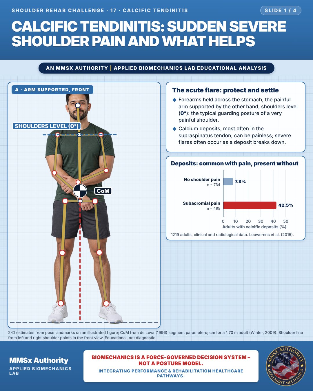
The acute flare: protect and settle
Forearms held across the stomach, the painful arm supported by the other hand, shoulders level (0°): the typical guarding posture of a very painful shoulder.
Calcium deposits, most often in the supraspinatus tendon, can be painless; severe flares often occur as a deposit breaks down.
Deposits: common with pain, present without
Movement B: Pendulum circles (side view)
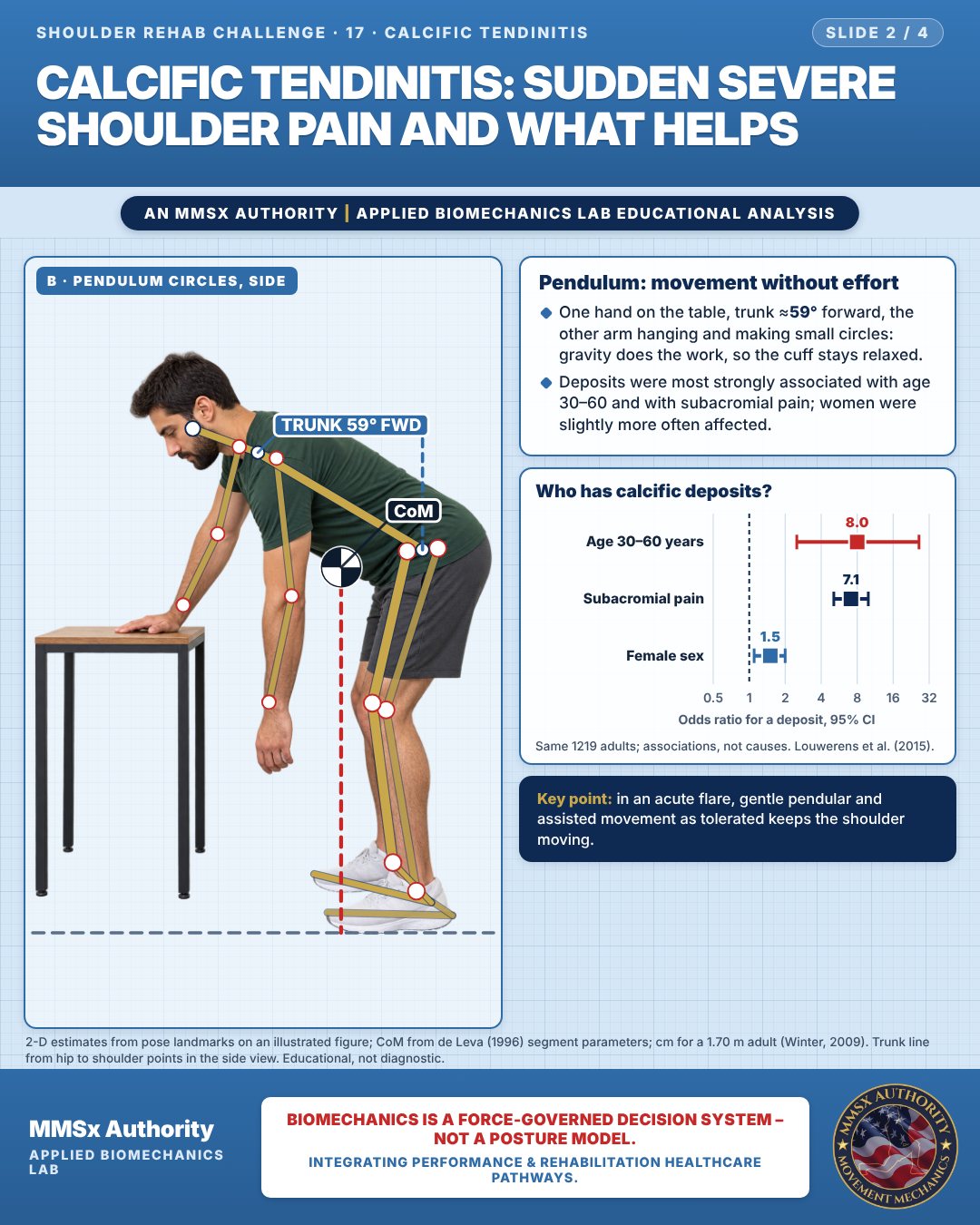
Pendulum: movement without effort
One hand on the table, trunk ≈59° forward, the other arm hanging and making small circles: gravity does the work, so the cuff stays relaxed.
Deposits were most strongly associated with age 30–60 and with subacromial pain; women were slightly more often affected.
Who has calcific deposits?
Movement C: Assisted arm raise (side view)
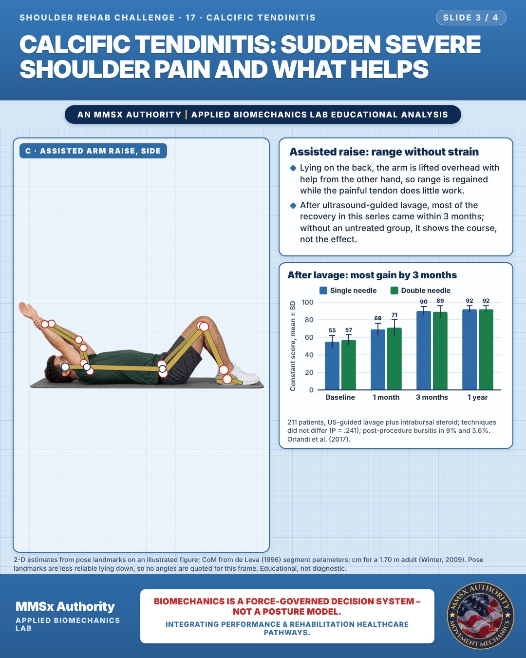
Assisted raise: range without strain
Lying on the back, the arm is lifted overhead with help from the other hand, so range is regained while the painful tendon does little work.
After ultrasound-guided lavage, most of the recovery in this series came within 3 months; without an untreated group, it shows the course, not the effect.
After lavage: most gain by 3 months
Decision pathway
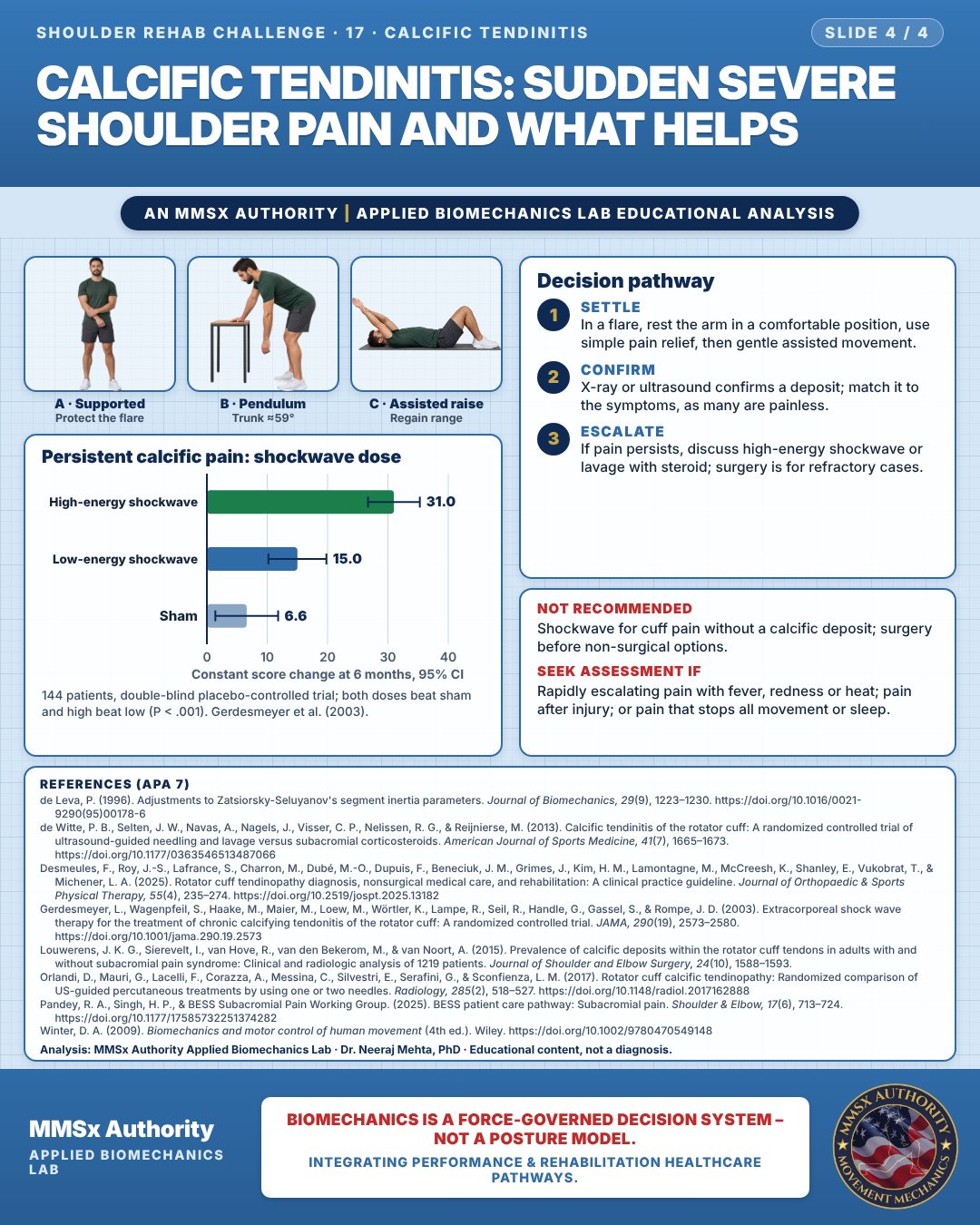
Persistent calcific pain: shockwave dose
Settle
In a flare, rest the arm in a comfortable position, use simple pain relief, then gentle assisted movement.
Confirm
X-ray or ultrasound confirms a deposit; match it to the symptoms, as many are painless.
Escalate
If pain persists, discuss high-energy shockwave or lavage with steroid; surgery is for refractory cases.
What the evidence supports
- Persistent symptomatic deposits after conservative care: options with randomised evidence are high-energy ESWT (Gerdesmeyer et al., 2003) and ultrasound-guided lavage plus subacromial steroid (de Witte et al., 2013). Surgery is reserved for refractory cases.
Myth and evidence
Myth“A calcium deposit on the X-ray always explains the pain and must be removed.”
EvidenceAbout 1 in 13 pain-free adults has deposits (7.8%; Louwerens et al., 2015). When deposits are symptomatic and persistent, non-surgical options such as ESWT and lavage have randomised evidence (Gerdesmeyer et al., 2003; de Witte et al., 2013).
Cautions
Not recommended
Shockwave for cuff pain without a calcific deposit; surgery before non-surgical options.
Seek assessment if
Rapidly escalating pain with fever, redness or heat; pain after injury; or pain that stops all movement or sleep.
Methods note
2-D estimates from pose landmarks on an illustrated figure; CoM from de Leva (1996) segment parameters; cm for a 1.70 m adult (Winter, 2009). Shoulder line from left and right shoulder points in the front view. Trunk line from hip to shoulder points in the side view. Pose landmarks are less reliable lying down, so no angles are quoted for this frame. These are educational estimates, not a diagnosis.
References
- de Leva, P. (1996). Adjustments to Zatsiorsky-Seluyanov's segment inertia parameters. Journal of Biomechanics, 29(9), 1223–1230. https://doi.org/10.1016/0021-9290(95)00178-6
- de Witte, P. B., Selten, J. W., Navas, A., Nagels, J., Visser, C. P., Nelissen, R. G., & Reijnierse, M. (2013). Calcific tendinitis of the rotator cuff: A randomized controlled trial of ultrasound-guided needling and lavage versus subacromial corticosteroids. American Journal of Sports Medicine, 41(7), 1665–1673. https://doi.org/10.1177/0363546513487066
- Desmeules, F., Roy, J.-S., Lafrance, S., Charron, M., Dubé, M.-O., Dupuis, F., Beneciuk, J. M., Grimes, J., Kim, H. M., Lamontagne, M., McCreesh, K., Shanley, E., Vukobrat, T., & Michener, L. A. (2025). Rotator cuff tendinopathy diagnosis, nonsurgical medical care, and rehabilitation: A clinical practice guideline. Journal of Orthopaedic & Sports Physical Therapy, 55(4), 235–274. https://doi.org/10.2519/jospt.2025.13182
- Gerdesmeyer, L., Wagenpfeil, S., Haake, M., Maier, M., Loew, M., Wörtler, K., Lampe, R., Seil, R., Handle, G., Gassel, S., & Rompe, J. D. (2003). Extracorporeal shock wave therapy for the treatment of chronic calcifying tendonitis of the rotator cuff: A randomized controlled trial. JAMA, 290(19), 2573–2580. https://doi.org/10.1001/jama.290.19.2573
- Louwerens, J. K. G., Sierevelt, I., van Hove, R., van den Bekerom, M., & van Noort, A. (2015). Prevalence of calcific deposits within the rotator cuff tendons in adults with and without subacromial pain syndrome: Clinical and radiologic analysis of 1219 patients. Journal of Shoulder and Elbow Surgery, 24(10), 1588–1593.
- Orlandi, D., Mauri, G., Lacelli, F., Corazza, A., Messina, C., Silvestri, E., Serafini, G., & Sconfienza, L. M. (2017). Rotator cuff calcific tendinopathy: Randomized comparison of US-guided percutaneous treatments by using one or two needles. Radiology, 285(2), 518–527. https://doi.org/10.1148/radiol.2017162888
- Pandey, R. A., Singh, H. P., & BESS Subacromial Pain Working Group. (2025). BESS patient care pathway: Subacromial pain. Shoulder & Elbow, 17(6), 713–724. https://doi.org/10.1177/17585732251374282
- Winter, D. A. (2009). Biomechanics and motor control of human movement (4th ed.). Wiley. https://doi.org/10.1002/9780470549148
Cite this article
Mehta, N. (2026, October 24). Calcific tendinitis: sudden severe shoulder pain and what helps. MMSx Authority Applied Biomechanics Lab. https://mmsxauthority.org/edu/analysis/shoulder/17-calcific-tendinitis-sudden-severe-shoulder-pain-and-what-helps/
Part of the collection
Mehta, N. (2026). Shoulder Rehabilitation: 50 Evidence-Based Biomechanics Analyses [Collection]. MMSx Authority Applied Biomechanics Lab. Zenodo. https://doi.org/10.5281/zenodo.23735725
All Shoulder Rehab Challenge articles · Rehabilitation Library