Shoulder Rehab Challenge · 06
Rotator cuff tears on MRI: why many painless shoulders have them too
Rotator cuff tears and other cuff abnormalities are common imaging findings in people with no shoulder symptoms, and their frequency rises steeply with age.
Key points
- Examine first. Subacromial pain is a clinical diagnosis; imaging is not needed to start treatment.
- Match. Interpret any scan alongside the examination; a tear may be a normal age change.
- Image if needed. After 12 weeks without progress, ultrasound is preferred; X-ray after trauma or with red flags.
What it is
Rotator cuff tears and other cuff abnormalities are common imaging findings in people with no shoulder symptoms, and their frequency rises steeply with age. An imaging finding therefore needs to be matched to the clinical picture before it is treated as the cause of pain.
How we analysed the movement
Three movements relevant to this topic were analysed from illustrated reference figures. Joint centres were located with two-dimensional pose estimation; arm elevation and elbow angles, shoulder level, the whole-body centre of mass and, where relevant, the arm lever (the horizontal distance from the shoulder joint to the centre of mass of the arm, with any hand load) and the resulting shoulder moment were then calculated from standard segment parameters. The figures are educational estimates that show how position changes mechanical demand; they are not measurements of a patient. Every research value quoted below comes from the published source cited beside it.
Movement A: Both arms overhead (front view)
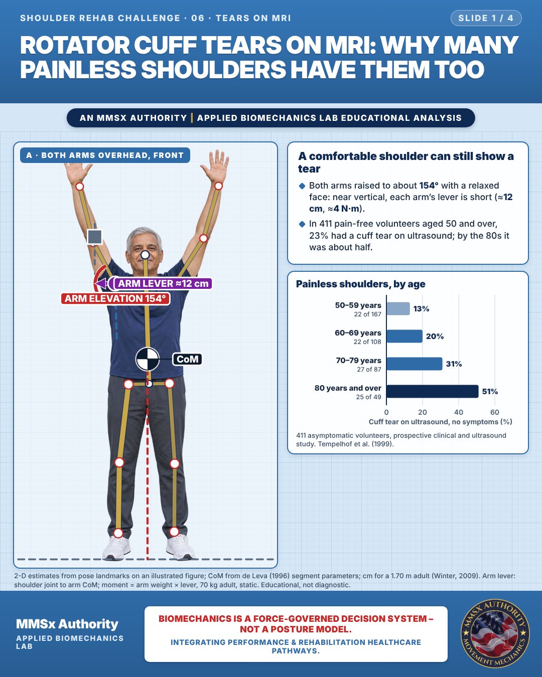
A comfortable shoulder can still show a tear
Both arms raised to about 154° with a relaxed face: near vertical, each arm’s lever is short (≈12 cm, ≈4 N·m).
In 411 pain-free volunteers aged 50 and over, 23% had a cuff tear on ultrasound; by the 80s it was about half.
Painless shoulders, by age
Movement B: Suitcase to rack (side view)
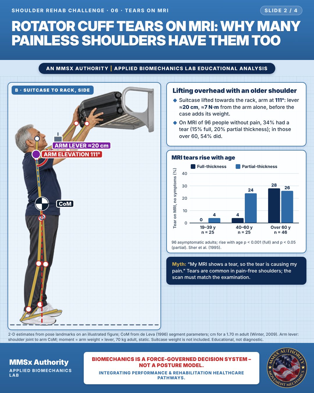
Lifting overhead with an older shoulder
Suitcase lifted towards the rack, arm at 111°: lever ≈20 cm, ≈7 N·m from the arm alone, before the case adds its weight.
On MRI of 96 people without pain, 34% had a tear (15% full, 20% partial thickness); in those over 60, 54% did.
MRI tears rise with age
Movement C: Wall push-up (side view)
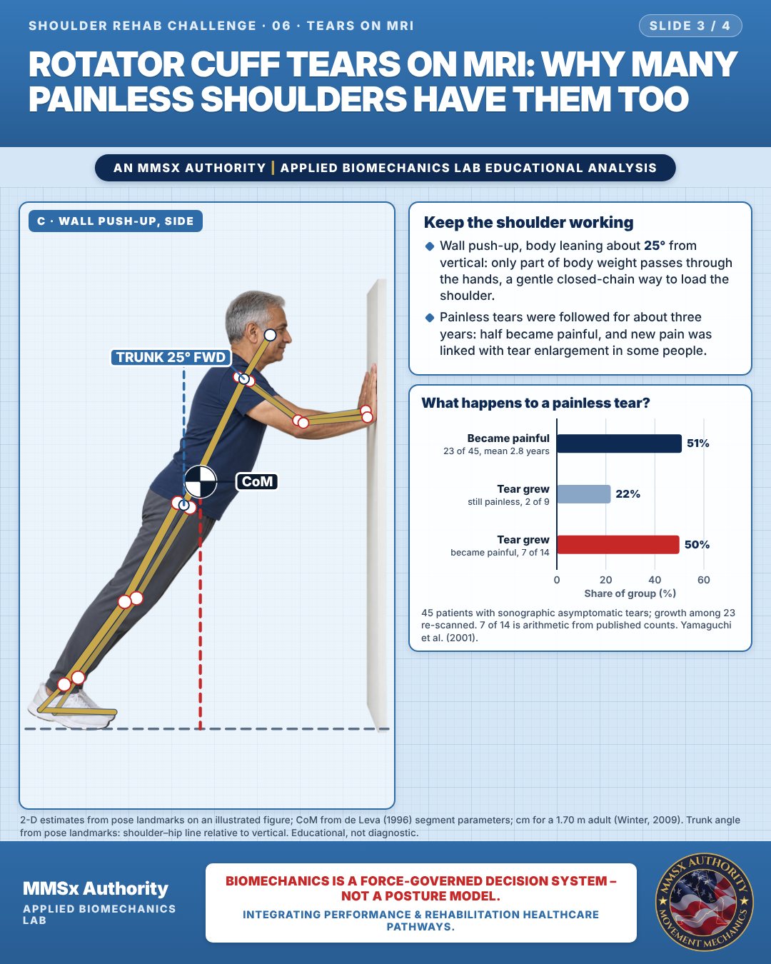
Keep the shoulder working
Wall push-up, body leaning about 25° from vertical: only part of body weight passes through the hands, a gentle closed-chain way to load the shoulder.
Painless tears were followed for about three years: half became painful, and new pain was linked with tear enlargement in some people.
What happens to a painless tear?
Decision pathway
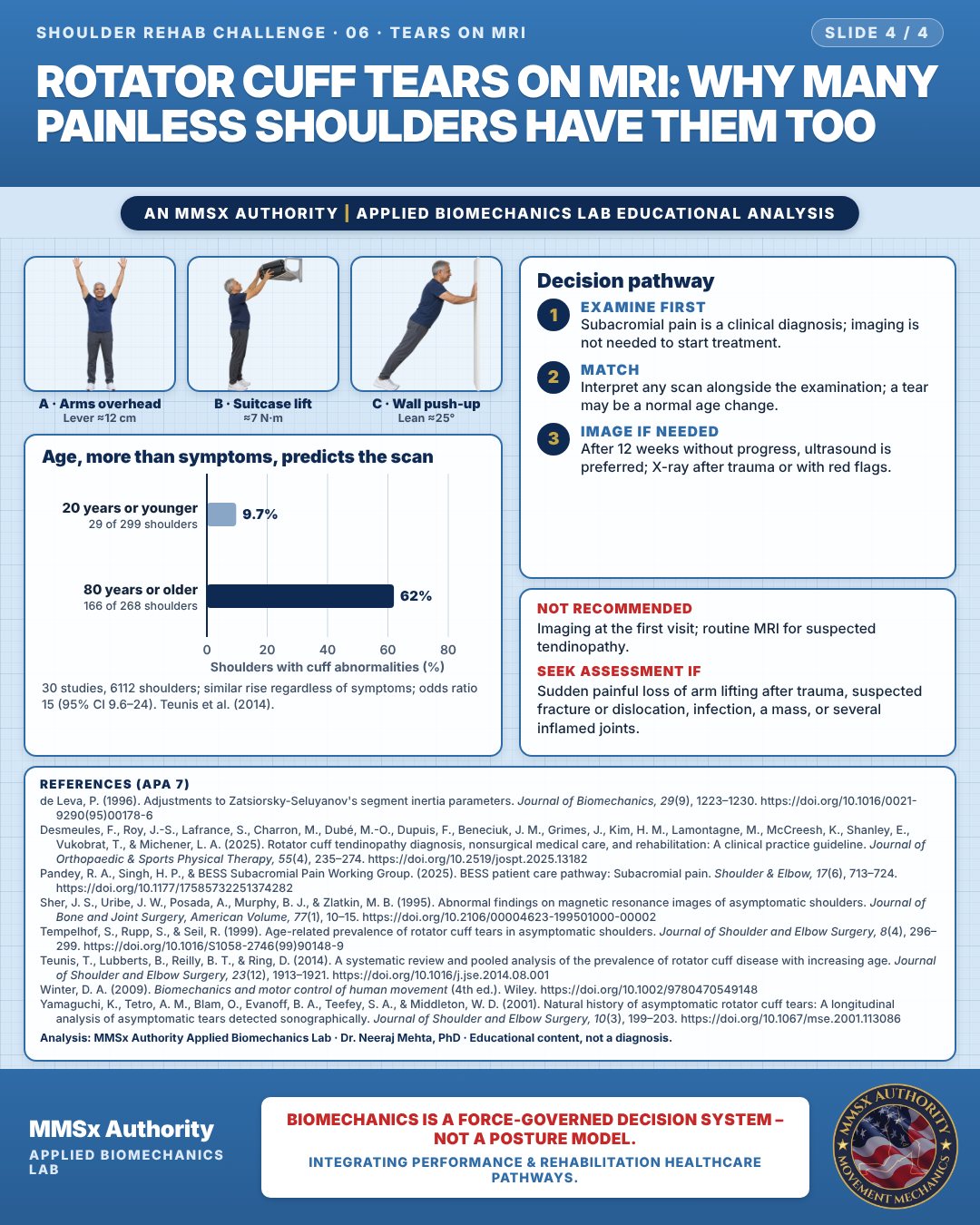
Age, more than symptoms, predicts the scan
Examine first
Subacromial pain is a clinical diagnosis; imaging is not needed to start treatment.
Match
Interpret any scan alongside the examination; a tear may be a normal age change.
Image if needed
After 12 weeks without progress, ultrasound is preferred; X-ray after trauma or with red flags.
What the evidence supports
- Imaging should not be used initially for suspected rotator cuff tendinopathy (Grade F). If imaging is needed after 12 weeks of failed conservative care, ultrasound is preferred, and MRI is not routinely recommended (Desmeules et al., 2025).
- BESS: subacromial pain is a clinical diagnosis and imaging is not essential to start treatment. Plain radiographs are advised after trauma, with red flags, with severe pain or stiffness, or for long-lasting symptoms. Ultrasound in experienced hands is a low-cost first-line option (Pandey et al., 2025).
Myth and evidence
Myth“My MRI shows a tear, so the tear must be causing my pain.”
EvidenceIn people with no pain, tears were found in 54% of over-60s on MRI (Sher et al., 1995) and in 51% of over-80s on ultrasound (Tempelhof et al., 1999). Abnormalities rise with age regardless of symptoms (Teunis et al., 2014). The scan must be interpreted alongside the clinical examination.
Cautions
Not recommended
Imaging at the first visit; routine MRI for suspected tendinopathy.
Seek assessment if
Sudden painful loss of arm lifting after trauma, suspected fracture or dislocation, infection, a mass, or several inflamed joints.
Methods note
2-D estimates from pose landmarks on an illustrated figure; CoM from de Leva (1996) segment parameters; cm for a 1.70 m adult (Winter, 2009). Arm lever: shoulder joint to arm CoM; moment = arm weight × lever, 70 kg adult, static. Suitcase weight is not included. Trunk angle from pose landmarks: shoulder–hip line relative to vertical. These are educational estimates, not a diagnosis.
References
- de Leva, P. (1996). Adjustments to Zatsiorsky-Seluyanov's segment inertia parameters. Journal of Biomechanics, 29(9), 1223–1230. https://doi.org/10.1016/0021-9290(95)00178-6
- Desmeules, F., Roy, J.-S., Lafrance, S., Charron, M., Dubé, M.-O., Dupuis, F., Beneciuk, J. M., Grimes, J., Kim, H. M., Lamontagne, M., McCreesh, K., Shanley, E., Vukobrat, T., & Michener, L. A. (2025). Rotator cuff tendinopathy diagnosis, nonsurgical medical care, and rehabilitation: A clinical practice guideline. Journal of Orthopaedic & Sports Physical Therapy, 55(4), 235–274. https://doi.org/10.2519/jospt.2025.13182
- Pandey, R. A., Singh, H. P., & BESS Subacromial Pain Working Group. (2025). BESS patient care pathway: Subacromial pain. Shoulder & Elbow, 17(6), 713–724. https://doi.org/10.1177/17585732251374282
- Sher, J. S., Uribe, J. W., Posada, A., Murphy, B. J., & Zlatkin, M. B. (1995). Abnormal findings on magnetic resonance images of asymptomatic shoulders. Journal of Bone and Joint Surgery, American Volume, 77(1), 10–15. https://doi.org/10.2106/00004623-199501000-00002
- Tempelhof, S., Rupp, S., & Seil, R. (1999). Age-related prevalence of rotator cuff tears in asymptomatic shoulders. Journal of Shoulder and Elbow Surgery, 8(4), 296–299. https://doi.org/10.1016/S1058-2746(99)90148-9
- Teunis, T., Lubberts, B., Reilly, B. T., & Ring, D. (2014). A systematic review and pooled analysis of the prevalence of rotator cuff disease with increasing age. Journal of Shoulder and Elbow Surgery, 23(12), 1913–1921. https://doi.org/10.1016/j.jse.2014.08.001
- Winter, D. A. (2009). Biomechanics and motor control of human movement (4th ed.). Wiley. https://doi.org/10.1002/9780470549148
- Yamaguchi, K., Tetro, A. M., Blam, O., Evanoff, B. A., Teefey, S. A., & Middleton, W. D. (2001). Natural history of asymptomatic rotator cuff tears: A longitudinal analysis of asymptomatic tears detected sonographically. Journal of Shoulder and Elbow Surgery, 10(3), 199–203. https://doi.org/10.1067/mse.2001.113086
Cite this article
Mehta, N. (2026, October 24). Rotator cuff tears on MRI: why many painless shoulders have them too. MMSx Authority Applied Biomechanics Lab. https://mmsxauthority.org/edu/analysis/shoulder/06-rotator-cuff-tears-on-mri-why-many-painless-shoulders-have-them-too/
Part of the collection
Mehta, N. (2026). Shoulder Rehabilitation: 50 Evidence-Based Biomechanics Analyses [Collection]. MMSx Authority Applied Biomechanics Lab. Zenodo. https://doi.org/10.5281/zenodo.23735725
All Shoulder Rehab Challenge articles · Rehabilitation Library