Shoulder Rehab Challenge · 03
Subacromial pain (impingement): why the old "pinching" explanation is changing
Subacromial pain (formerly "subacromial impingement syndrome") is shoulder pain, typically lateral and provoked by arm elevation, that is attributed…
Key points
- Explain. Subacromial pain is a clinical diagnosis; reassure that narrowing with arm elevation is normal.
- Load. Specific, progressive exercise is first line; a single injection can be considered if no better by 6–8 weeks.
- Review. Refer if no improvement by 12 weeks; surgery only after six months of non-surgical care.
What it is
Subacromial pain (formerly "subacromial impingement syndrome") is shoulder pain, typically lateral and provoked by arm elevation, that is attributed to the rotator cuff and subacromial bursa after other causes have been excluded. It is a clinical diagnosis (Pandey et al., 2025). The idea that it is caused purely by mechanical pinching under the acromion is no longer well supported, because removing bone (decompression) did not beat placebo surgery (Beard et al., 2018).
How we analysed the movement
Three movements relevant to this topic were analysed from illustrated reference figures. Joint centres were located with two-dimensional pose estimation; arm elevation and elbow angles, shoulder level, the whole-body centre of mass and, where relevant, the arm lever (the horizontal distance from the shoulder joint to the centre of mass of the arm, with any hand load) and the resulting shoulder moment were then calculated from standard segment parameters. The figures are educational estimates that show how position changes mechanical demand; they are not measurements of a patient. Every research value quoted below comes from the published source cited beside it.
Movement A: Scaption raise (front view)
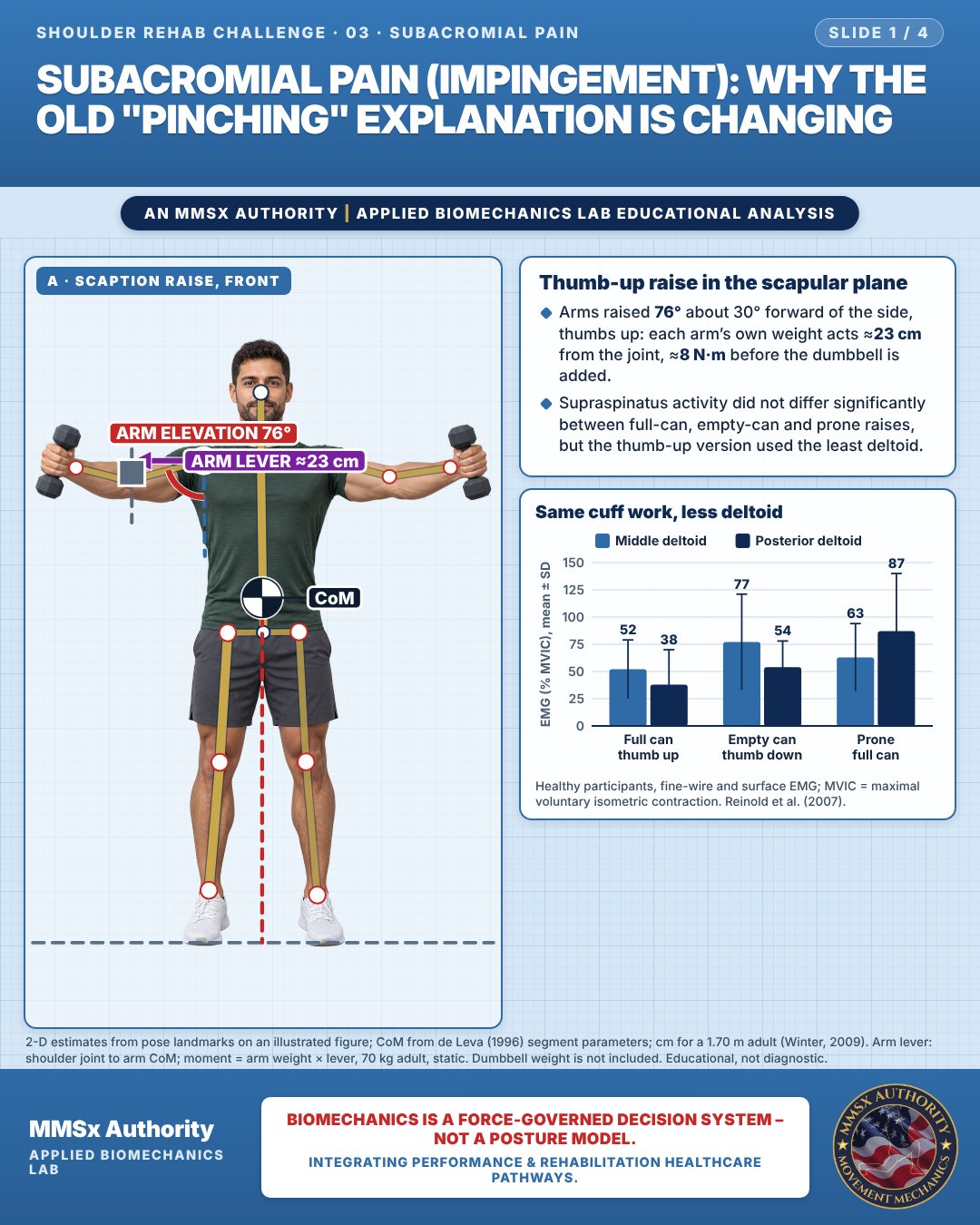
Thumb-up raise in the scapular plane
Arms raised 76° about 30° forward of the side, thumbs up: each arm’s own weight acts ≈23 cm from the joint, ≈8 N·m before the dumbbell is added.
Supraspinatus activity did not differ significantly between full-can, empty-can and prone raises, but the thumb-up version used the least deltoid.
Same cuff work, less deltoid
Movement B: Hanging a shirt (side view)
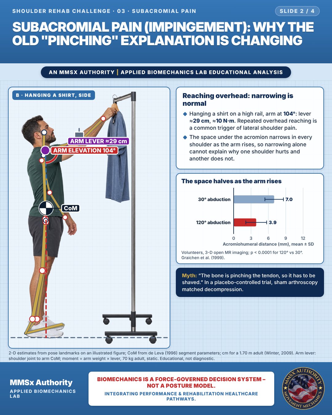
Reaching overhead: narrowing is normal
Hanging a shirt on a high rail, arm at 104°: lever ≈29 cm, ≈10 N·m. Repeated overhead reaching is a common trigger of lateral shoulder pain.
The space under the acromion narrows in every shoulder as the arm rises, so narrowing alone cannot explain why one shoulder hurts and another does not.
The space halves as the arm rises
Movement C: Side-lying rotation (front view)
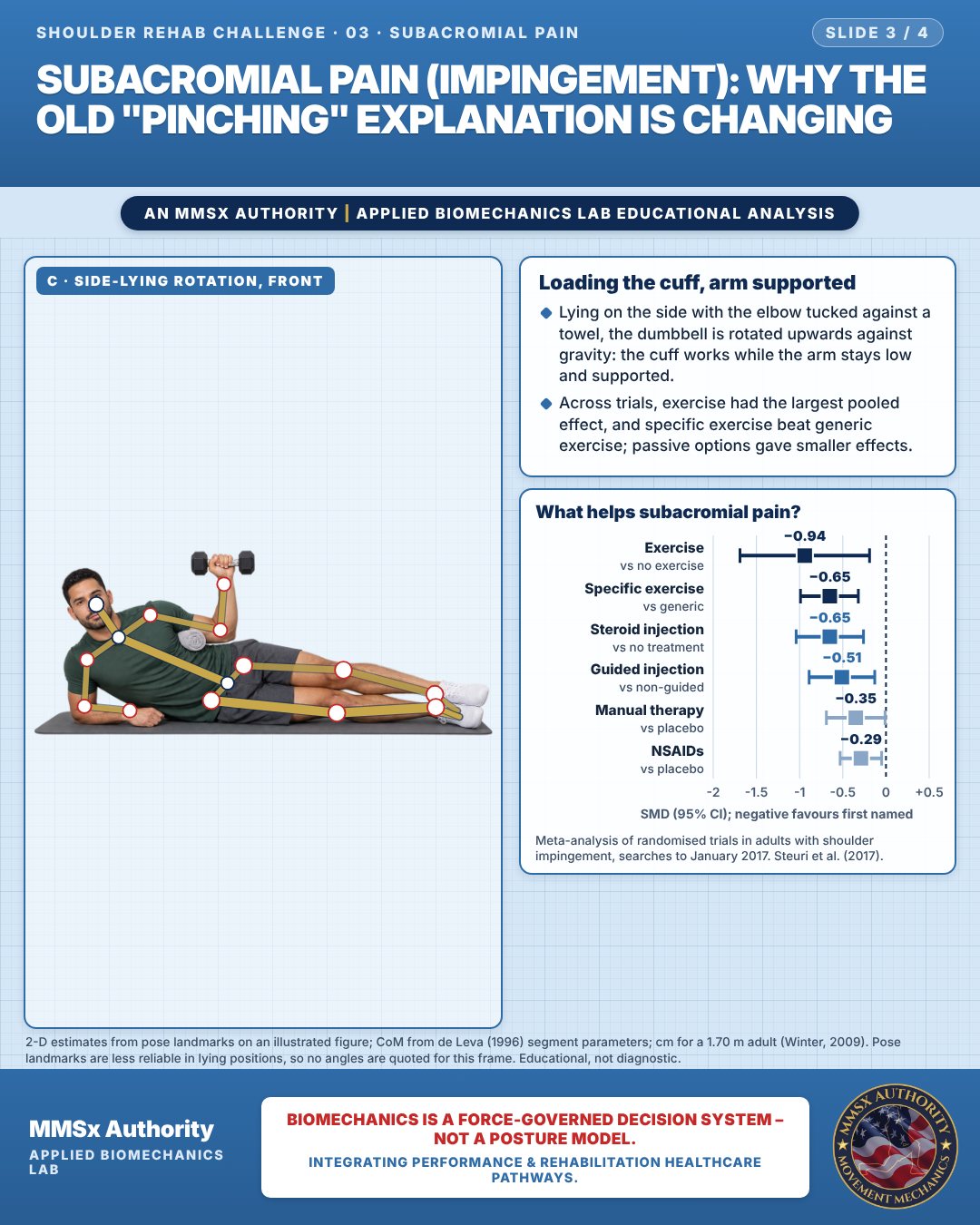
Loading the cuff, arm supported
Lying on the side with the elbow tucked against a towel, the dumbbell is rotated upwards against gravity: the cuff works while the arm stays low and supported.
Across trials, exercise had the largest pooled effect, and specific exercise beat generic exercise; passive options gave smaller effects.
What helps subacromial pain?
Decision pathway
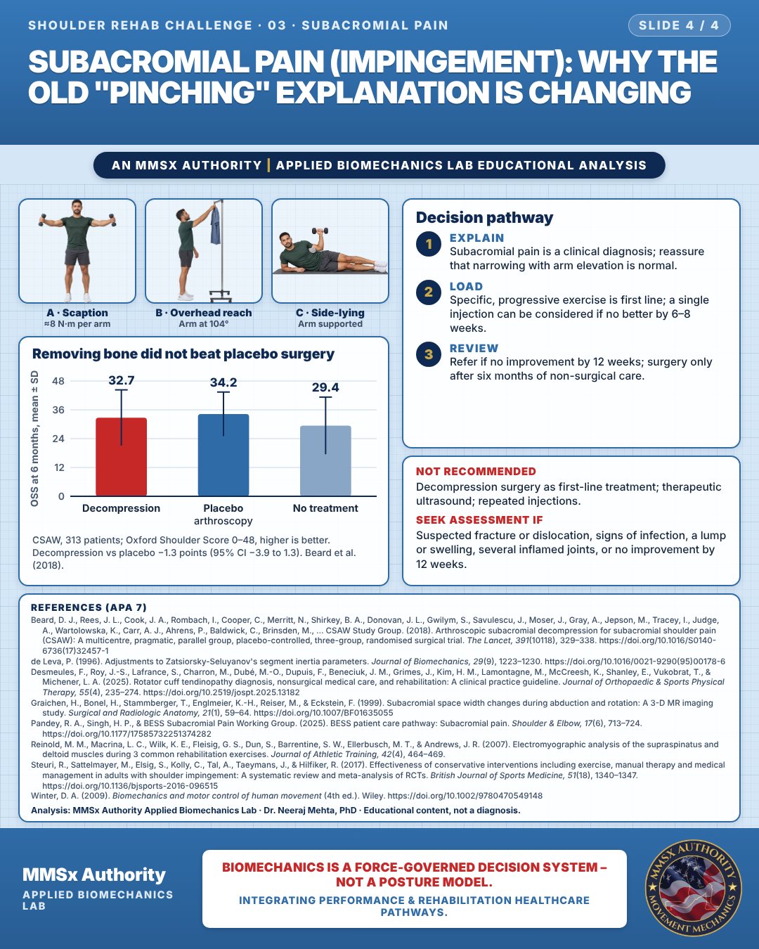
Removing bone did not beat placebo surgery
Explain
Subacromial pain is a clinical diagnosis; reassure that narrowing with arm elevation is normal.
Load
Specific, progressive exercise is first line; a single injection can be considered if no better by 6–8 weeks.
Review
Refer if no improvement by 12 weeks; surgery only after six months of non-surgical care.
What the evidence supports
- First line: education and progressive exercise therapy (Grade A for exercise) (Desmeules et al., 2025). Specific exercise outperforms generic exercise (Steuri et al., 2017).
- A single subacromial steroid injection can be considered, after shared decision-making, if there is no improvement by 6–8 weeks; multiple injections showed no added benefit (Pandey et al., 2025).
Myth and evidence
Myth“The bone is pinching the tendon, so it has to be shaved.”
EvidencePlacebo arthroscopy matched decompression surgery (Beard et al., 2018). Narrowing of the subacromial space is a normal feature of arm elevation (Graichen et al., 1999). Exercise has the largest pooled benefit (Steuri et al., 2017).
Cautions
Not recommended
Decompression surgery as first-line treatment; therapeutic ultrasound; repeated injections.
Seek assessment if
Suspected fracture or dislocation, signs of infection, a lump or swelling, several inflamed joints, or no improvement by 12 weeks.
Methods note
2-D estimates from pose landmarks on an illustrated figure; CoM from de Leva (1996) segment parameters; cm for a 1.70 m adult (Winter, 2009). Arm lever: shoulder joint to arm CoM; moment = arm weight × lever, 70 kg adult, static. Dumbbell weight is not included. Pose landmarks are less reliable in lying positions, so no angles are quoted for this frame. These are educational estimates, not a diagnosis.
References
- Beard, D. J., Rees, J. L., Cook, J. A., Rombach, I., Cooper, C., Merritt, N., Shirkey, B. A., Donovan, J. L., Gwilym, S., Savulescu, J., Moser, J., Gray, A., Jepson, M., Tracey, I., Judge, A., Wartolowska, K., Carr, A. J., Ahrens, P., Baldwick, C., Brinsden, M., … CSAW Study Group. (2018). Arthroscopic subacromial decompression for subacromial shoulder pain (CSAW): A multicentre, pragmatic, parallel group, placebo-controlled, three-group, randomised surgical trial. The Lancet, 391(10118), 329–338. https://doi.org/10.1016/S0140-6736(17)32457-1
- de Leva, P. (1996). Adjustments to Zatsiorsky-Seluyanov's segment inertia parameters. Journal of Biomechanics, 29(9), 1223–1230. https://doi.org/10.1016/0021-9290(95)00178-6
- Desmeules, F., Roy, J.-S., Lafrance, S., Charron, M., Dubé, M.-O., Dupuis, F., Beneciuk, J. M., Grimes, J., Kim, H. M., Lamontagne, M., McCreesh, K., Shanley, E., Vukobrat, T., & Michener, L. A. (2025). Rotator cuff tendinopathy diagnosis, nonsurgical medical care, and rehabilitation: A clinical practice guideline. Journal of Orthopaedic & Sports Physical Therapy, 55(4), 235–274. https://doi.org/10.2519/jospt.2025.13182
- Graichen, H., Bonel, H., Stammberger, T., Englmeier, K.-H., Reiser, M., & Eckstein, F. (1999). Subacromial space width changes during abduction and rotation: A 3-D MR imaging study. Surgical and Radiologic Anatomy, 21(1), 59–64. https://doi.org/10.1007/BF01635055
- Pandey, R. A., Singh, H. P., & BESS Subacromial Pain Working Group. (2025). BESS patient care pathway: Subacromial pain. Shoulder & Elbow, 17(6), 713–724. https://doi.org/10.1177/17585732251374282
- Reinold, M. M., Macrina, L. C., Wilk, K. E., Fleisig, G. S., Dun, S., Barrentine, S. W., Ellerbusch, M. T., & Andrews, J. R. (2007). Electromyographic analysis of the supraspinatus and deltoid muscles during 3 common rehabilitation exercises. Journal of Athletic Training, 42(4), 464–469.
- Steuri, R., Sattelmayer, M., Elsig, S., Kolly, C., Tal, A., Taeymans, J., & Hilfiker, R. (2017). Effectiveness of conservative interventions including exercise, manual therapy and medical management in adults with shoulder impingement: A systematic review and meta-analysis of RCTs. British Journal of Sports Medicine, 51(18), 1340–1347. https://doi.org/10.1136/bjsports-2016-096515
- Winter, D. A. (2009). Biomechanics and motor control of human movement (4th ed.). Wiley. https://doi.org/10.1002/9780470549148
Cite this article
Mehta, N. (2026, October 24). Subacromial pain (impingement): why the old "pinching" explanation is changing. MMSx Authority Applied Biomechanics Lab. https://mmsxauthority.org/edu/analysis/shoulder/03-subacromial-pain-impingement-why-the-old-pinching-explanation-is/
Part of the collection
Mehta, N. (2026). Shoulder Rehabilitation: 50 Evidence-Based Biomechanics Analyses [Collection]. MMSx Authority Applied Biomechanics Lab. Zenodo. https://doi.org/10.5281/zenodo.23735725
All Shoulder Rehab Challenge articles · Rehabilitation Library