Shoulder Rehab Challenge · 02
Shoulder pain when lifting your arm: what the painful arc tells us
A painful arc is pain felt during part of active arm elevation, classically mid-range abduction, that eases above or below that range.
Key points
- Assess. Take a full history and screen for red flags; combine the arc with other tests rather than relying on one sign.
- Load. Start progressive exercise; grade the lever with a bent elbow, a smaller arc and lighter weights.
- Review. Consider a single injection if no better by 6–8 weeks; refer if no improvement by 12 weeks.
What it is
A painful arc is pain felt during part of active arm elevation, classically mid-range abduction, that eases above or below that range. It is a clinical sign that raises suspicion of a subacromial or rotator cuff source. It is not a diagnosis of a specific structural lesion.
How we analysed the movement
Three movements relevant to this topic were analysed from illustrated reference figures. Joint centres were located with two-dimensional pose estimation; arm elevation and elbow angles, shoulder level, the whole-body centre of mass and, where relevant, the arm lever (the horizontal distance from the shoulder joint to the centre of mass of the arm, with any hand load) and the resulting shoulder moment were then calculated from standard segment parameters. The figures are educational estimates that show how position changes mechanical demand; they are not measurements of a patient. Every research value quoted below comes from the published source cited beside it.
Movement A: Low arc (front view)
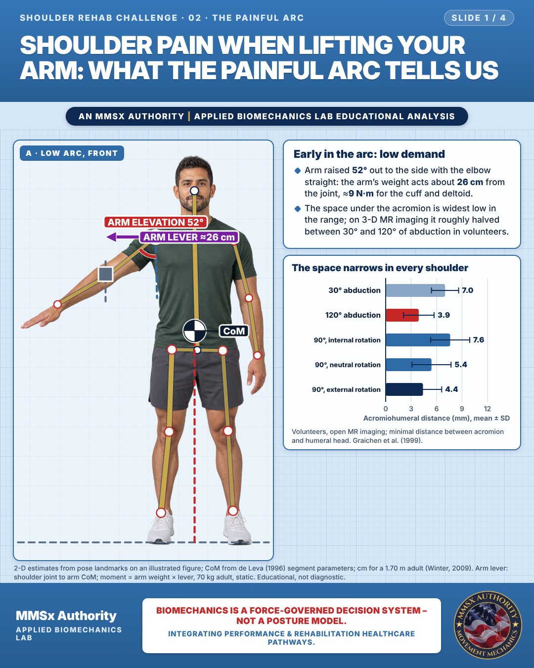
Early in the arc: low demand
Arm raised 52° out to the side with the elbow straight: the arm’s weight acts about 26 cm from the joint, ≈9 N·m for the cuff and deltoid.
The space under the acromion is widest low in the range; on 3-D MR imaging it roughly halved between 30° and 120° of abduction in volunteers.
The space narrows in every shoulder
Movement B: Shoulder height (front view)
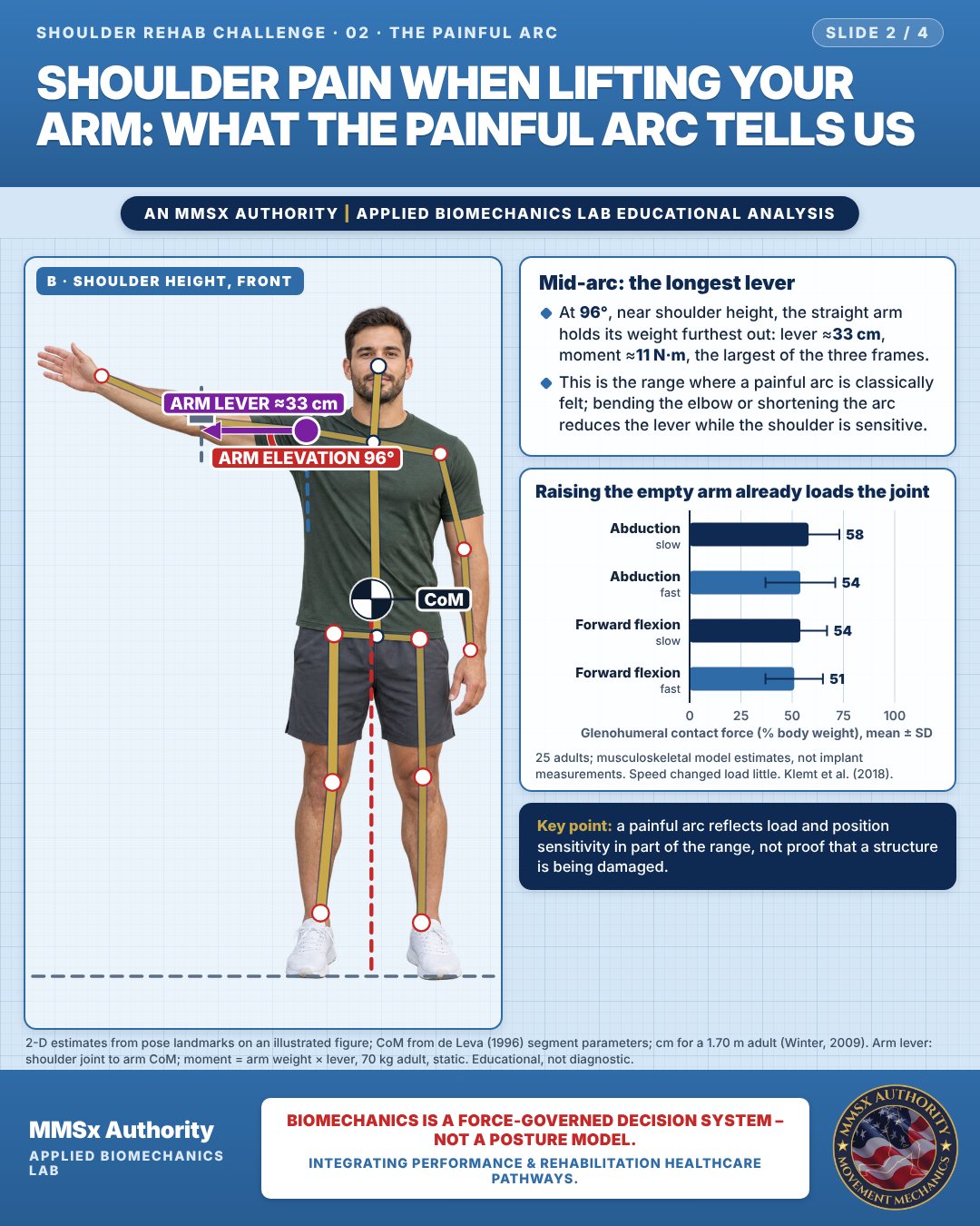
Mid-arc: the longest lever
At 96°, near shoulder height, the straight arm holds its weight furthest out: lever ≈33 cm, moment ≈11 N·m, the largest of the three frames.
This is the range where a painful arc is classically felt; bending the elbow or shortening the arc reduces the lever while the shoulder is sensitive.
Raising the empty arm already loads the joint
Movement C: Arm overhead (front view)
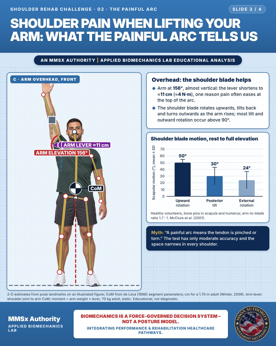
Overhead: the shoulder blade helps
Arm at 156°, almost vertical: the lever shortens to ≈11 cm (≈4 N·m), one reason pain often eases at the top of the arc.
The shoulder blade rotates upwards, tilts back and turns outwards as the arm rises; most tilt and outward rotation occur above 90°.
Shoulder blade motion, rest to full elevation
Decision pathway
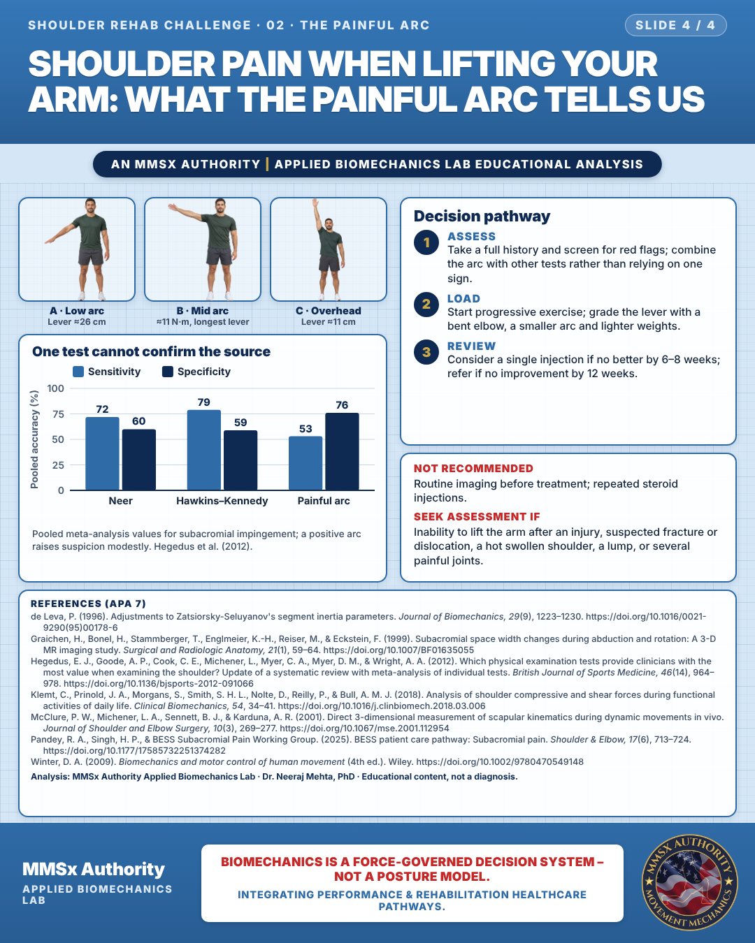
One test cannot confirm the source
Assess
Take a full history and screen for red flags; combine the arc with other tests rather than relying on one sign.
Load
Start progressive exercise; grade the lever with a bent elbow, a smaller arc and lighter weights.
Review
Consider a single injection if no better by 6–8 weeks; refer if no improvement by 12 weeks.
What the evidence supports
- BESS: consider a single subacromial steroid injection, after shared decision-making, if there is no improvement by 6–8 weeks; multiple injections showed no added benefit over a single one. Refer if there is no improvement by 12 weeks (Pandey et al., 2025).
Cautions
Not recommended
Routine imaging before treatment; repeated steroid injections.
Seek assessment if
Inability to lift the arm after an injury, suspected fracture or dislocation, a hot swollen shoulder, a lump, or several painful joints.
Methods note
2-D estimates from pose landmarks on an illustrated figure; CoM from de Leva (1996) segment parameters; cm for a 1.70 m adult (Winter, 2009). Arm lever: shoulder joint to arm CoM; moment = arm weight × lever, 70 kg adult, static. These are educational estimates, not a diagnosis.
References
- de Leva, P. (1996). Adjustments to Zatsiorsky-Seluyanov's segment inertia parameters. Journal of Biomechanics, 29(9), 1223–1230. https://doi.org/10.1016/0021-9290(95)00178-6
- Graichen, H., Bonel, H., Stammberger, T., Englmeier, K.-H., Reiser, M., & Eckstein, F. (1999). Subacromial space width changes during abduction and rotation: A 3-D MR imaging study. Surgical and Radiologic Anatomy, 21(1), 59–64. https://doi.org/10.1007/BF01635055
- Hegedus, E. J., Goode, A. P., Cook, C. E., Michener, L., Myer, C. A., Myer, D. M., & Wright, A. A. (2012). Which physical examination tests provide clinicians with the most value when examining the shoulder? Update of a systematic review with meta-analysis of individual tests. British Journal of Sports Medicine, 46(14), 964–978. https://doi.org/10.1136/bjsports-2012-091066
- Klemt, C., Prinold, J. A., Morgans, S., Smith, S. H. L., Nolte, D., Reilly, P., & Bull, A. M. J. (2018). Analysis of shoulder compressive and shear forces during functional activities of daily life. Clinical Biomechanics, 54, 34–41. https://doi.org/10.1016/j.clinbiomech.2018.03.006
- McClure, P. W., Michener, L. A., Sennett, B. J., & Karduna, A. R. (2001). Direct 3-dimensional measurement of scapular kinematics during dynamic movements in vivo. Journal of Shoulder and Elbow Surgery, 10(3), 269–277. https://doi.org/10.1067/mse.2001.112954
- Pandey, R. A., Singh, H. P., & BESS Subacromial Pain Working Group. (2025). BESS patient care pathway: Subacromial pain. Shoulder & Elbow, 17(6), 713–724. https://doi.org/10.1177/17585732251374282
- Winter, D. A. (2009). Biomechanics and motor control of human movement (4th ed.). Wiley. https://doi.org/10.1002/9780470549148
Cite this article
Mehta, N. (2026, October 24). Shoulder pain when lifting your arm: what the painful arc tells us. MMSx Authority Applied Biomechanics Lab. https://mmsxauthority.org/edu/analysis/shoulder/02-shoulder-pain-when-lifting-your-arm-what-the-painful-arc-tells-us/
Part of the collection
Mehta, N. (2026). Shoulder Rehabilitation: 50 Evidence-Based Biomechanics Analyses [Collection]. MMSx Authority Applied Biomechanics Lab. Zenodo. https://doi.org/10.5281/zenodo.23735725
All Shoulder Rehab Challenge articles · Rehabilitation Library