Lower Back Rehab Challenge · 17
Sacroiliac joint pain: why pain location alone cannot confirm the diagnosis
Sacroiliac joint (SIJ) pain is pain arising from the SIJ complex, typically felt in the buttock below L5, around the posterior superior iliac spine,…
Key points
- Rule out the spine. Test repeated lumbar movements first; centralising symptoms point away from the SIJ.
- Cluster the tests. Use three or more provocation tests together; never pain location or palpation alone.
- Treat the person. Graded, active loading of the lumbopelvic region guided by function; blocks only where they change care.
What it is
Sacroiliac joint (SIJ) pain is pain arising from the SIJ complex, typically felt in the buttock below L5, around the posterior superior iliac spine, sometimes referring into the thigh. Because pain in this area can also come from the lumbar spine, hip or other structures, the research reference standard is relief after a fluoroscopically guided anaesthetic block of the joint; clinically, the diagnosis is a probability built from several findings, not from pain location alone.
How we analysed the movement
Three movements relevant to this topic were analysed from illustrated reference figures. Joint centres were located with two-dimensional pose estimation; joint angles, the whole-body centre of mass and, where relevant, the low-back lever (the horizontal distance from the upper-body centre of mass to the L5/S1 joint) were then calculated from standard segment parameters. The figures are educational estimates that show how position changes mechanical demand; they are not measurements of a patient. Every research value quoted below comes from the published source cited beside it.
Movement A: Pointing to the SIJ (rear view)
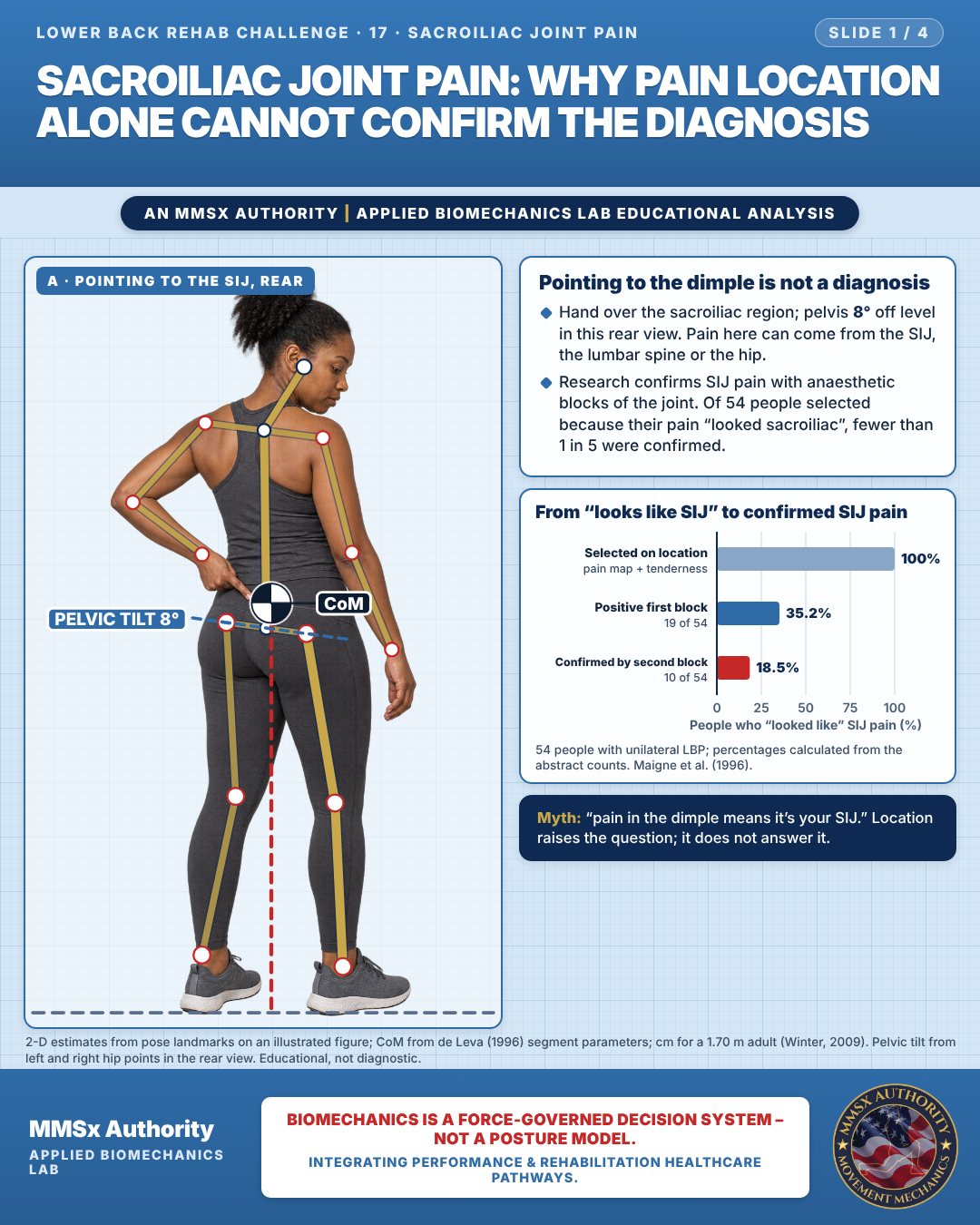
Pointing to the dimple is not a diagnosis
Hand over the sacroiliac region; pelvis 8° off level in this rear view. Pain here can come from the SIJ, the lumbar spine or the hip.
Research confirms SIJ pain with anaesthetic blocks of the joint. Of 54 people selected because their pain “looked sacroiliac”, fewer than 1 in 5 were confirmed.
From “looks like SIJ” to confirmed SIJ pain
Movement B: Single-leg step
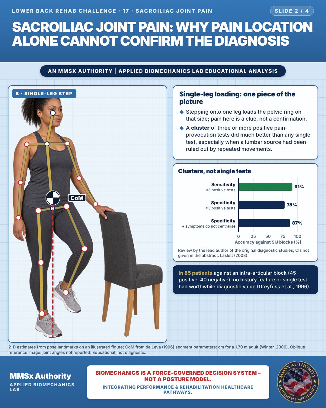
Single-leg loading: one piece of the picture
Stepping onto one leg loads the pelvic ring on that side; pain here is a clue, not a confirmation.
A cluster of three or more positive pain-provocation tests did much better than any single test, especially when a lumbar source had been ruled out by repeated movements.
Clusters, not single tests
Movement C: Supine testing position
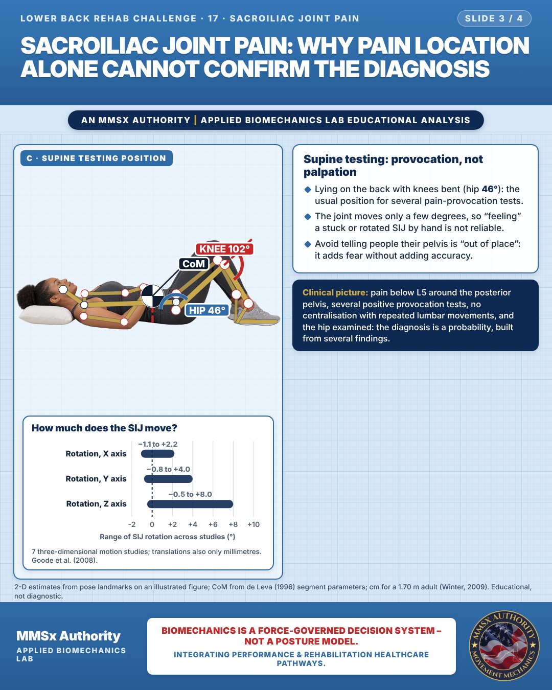
How much does the SIJ move?
Supine testing: provocation, not palpation
Lying on the back with knees bent (hip 46°): the usual position for several pain-provocation tests.
The joint moves only a few degrees, so “feeling” a stuck or rotated SIJ by hand is not reliable.
Avoid telling people their pelvis is “out of place”: it adds fear without adding accuracy.
Decision pathway
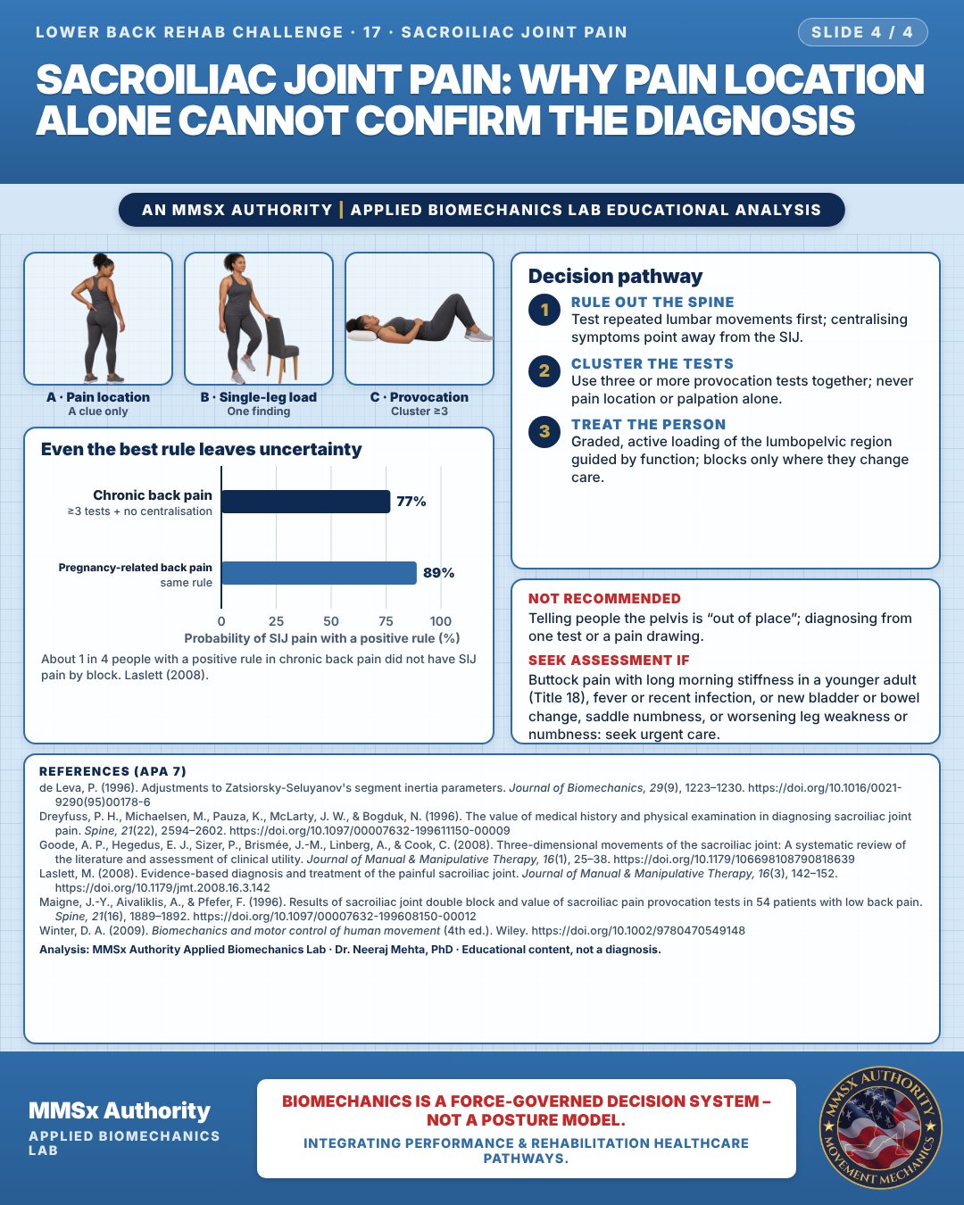
Even the best rule leaves uncertainty
Rule out the spine
Test repeated lumbar movements first; centralising symptoms point away from the SIJ.
Cluster the tests
Use three or more provocation tests together; never pain location or palpation alone.
Treat the person
Graded, active loading of the lumbopelvic region guided by function; blocks only where they change care.
What the evidence supports
- Use a cluster of provocation tests together with exclusion of a lumbar (centralising) source, rather than pain location or palpation of joint position (Laslett, 2008; Goode et al., 2008; Maigne et al., 1996; Dreyfuss et al., 1996).
Myth and evidence
Myth“A therapist can feel your SIJ is stuck or out of alignment.”
EvidenceSIJ rotations are only a few degrees (e.g. −1.1 to 2.2° about X) and palpation-based diagnosis has limited utility (Goode et al., 2008).
Cautions
Not recommended
Telling people the pelvis is “out of place”; diagnosing from one test or a pain drawing.
Seek assessment if
Buttock pain with long morning stiffness in a younger adult (Title 18), fever or recent infection, or new bladder or bowel change, saddle numbness, or worsening leg weakness or numbness: seek urgent care.
Methods note
2-D estimates from pose landmarks on an illustrated figure; CoM from de Leva (1996) segment parameters; cm for a 1.70 m adult (Winter, 2009). Pelvic tilt from left and right hip points in the rear view. Oblique reference image: joint angles not reported. These are educational estimates, not a diagnosis.
References
- de Leva, P. (1996). Adjustments to Zatsiorsky-Seluyanov's segment inertia parameters. Journal of Biomechanics, 29(9), 1223–1230. https://doi.org/10.1016/0021-9290(95)00178-6
- Dreyfuss, P. H., Michaelsen, M., Pauza, K., McLarty, J. W., & Bogduk, N. (1996). The value of medical history and physical examination in diagnosing sacroiliac joint pain. Spine, 21(22), 2594–2602. https://doi.org/10.1097/00007632-199611150-00009
- Goode, A. P., Hegedus, E. J., Sizer, P., Brismée, J.-M., Linberg, A., & Cook, C. (2008). Three-dimensional movements of the sacroiliac joint: A systematic review of the literature and assessment of clinical utility. Journal of Manual & Manipulative Therapy, 16(1), 25–38. https://doi.org/10.1179/106698108790818639
- Laslett, M. (2008). Evidence-based diagnosis and treatment of the painful sacroiliac joint. Journal of Manual & Manipulative Therapy, 16(3), 142–152. https://doi.org/10.1179/jmt.2008.16.3.142
- Maigne, J.-Y., Aivaliklis, A., & Pfefer, F. (1996). Results of sacroiliac joint double block and value of sacroiliac pain provocation tests in 54 patients with low back pain. Spine, 21(16), 1889–1892. https://doi.org/10.1097/00007632-199608150-00012
- Winter, D. A. (2009). Biomechanics and motor control of human movement (4th ed.). Wiley. https://doi.org/10.1002/9780470549148
Cite this article
Mehta, N. (2026, October 23). Sacroiliac joint pain: why pain location alone cannot confirm the diagnosis. MMSx Authority Applied Biomechanics Lab. https://mmsxauthority.org/edu/analysis/lower-back/17-sacroiliac-joint-pain-why-pain-location-alone-cannot-confirm-the/
Part of the collection
Mehta, N. (2026). Lower Back Rehabilitation: 50 Evidence-Based Biomechanics Analyses [Collection]. MMSx Authority Applied Biomechanics Lab. Zenodo. https://doi.org/10.5281/zenodo.23735778
All Lower Back Rehab Challenge articles · Rehabilitation Library