Lower Back Rehab Challenge · 12
Disc bulge on MRI: what the finding means for your movement
A disc bulge is a broad-based, symmetrical extension of the disc margin beyond the edges of the vertebral end plates (typically involving a large…
Key points
- Interpret in context. Match any finding to symptoms and the examination; alone it does not explain pain.
- Grade the load. Bending, lifting and exercise are progressed by response, not restricted by the report.
- Know when it matters. Nerve-root signs that are not improving, progressive deficit or red flags make imaging relevant.
What it is
A disc bulge is a broad-based, symmetrical extension of the disc margin beyond the edges of the vertebral end plates (typically involving a large part of the disc circumference). It is a description of disc shape on imaging, not a diagnosis of the cause of pain; it is common in people without back pain and becomes more common with age.
How we analysed the movement
Three movements relevant to this topic were analysed from illustrated reference figures. Joint centres were located with two-dimensional pose estimation; joint angles, the whole-body centre of mass and, where relevant, the low-back lever (the horizontal distance from the upper-body centre of mass to the L5/S1 joint) were then calculated from standard segment parameters. The figures are educational estimates that show how position changes mechanical demand; they are not measurements of a patient. Every research value quoted below comes from the published source cited beside it.
Movement A: Reading the report (front view)
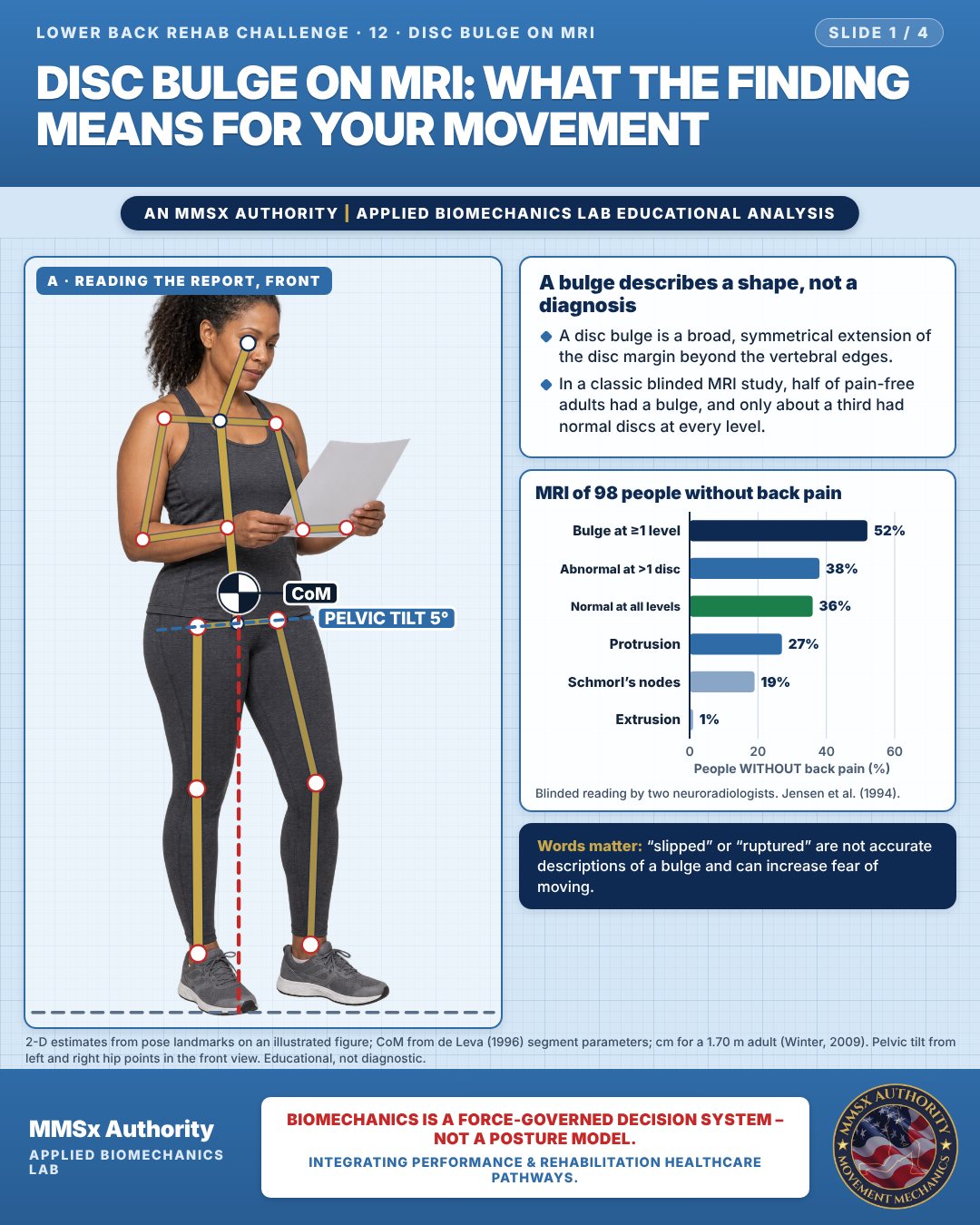
A bulge describes a shape, not a diagnosis
A disc bulge is a broad, symmetrical extension of the disc margin beyond the vertebral edges.
In a classic blinded MRI study, half of pain-free adults had a bulge, and only about a third had normal discs at every level.
MRI of 98 people without back pain
Movement B: Daily activity (front view)
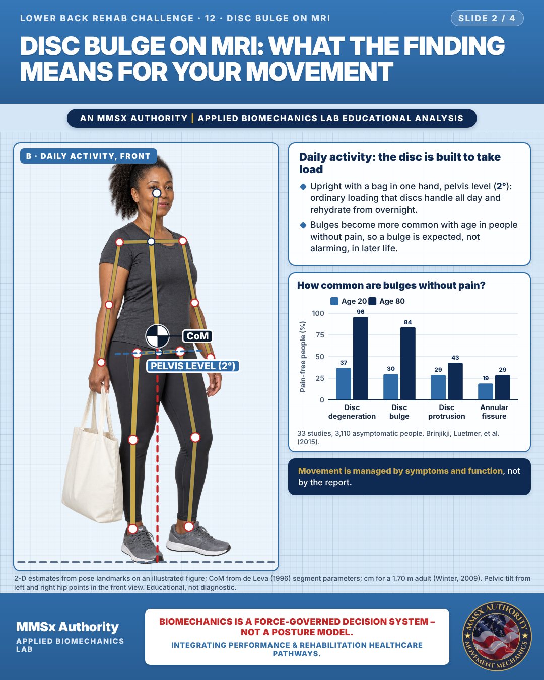
Daily activity: the disc is built to take load
Upright with a bag in one hand, pelvis level (2°): ordinary loading that discs handle all day and rehydrate from overnight.
Bulges become more common with age in people without pain, so a bulge is expected, not alarming, in later life.
How common are bulges without pain?
Movement C: Step-up (side view)
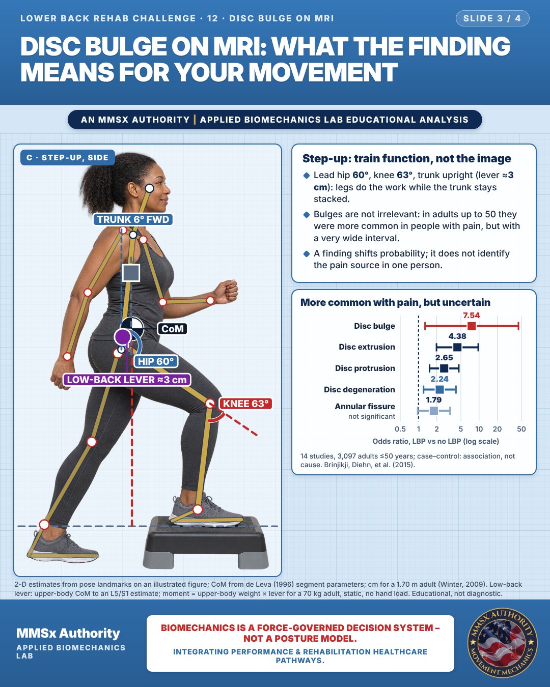
Step-up: train function, not the image
Lead hip 60°, knee 63°, trunk upright (lever ≈3 cm): legs do the work while the trunk stays stacked.
Bulges are not irrelevant: in adults up to 50 they were more common in people with pain, but with a very wide interval.
A finding shifts probability; it does not identify the pain source in one person.
More common with pain, but uncertain
Decision pathway
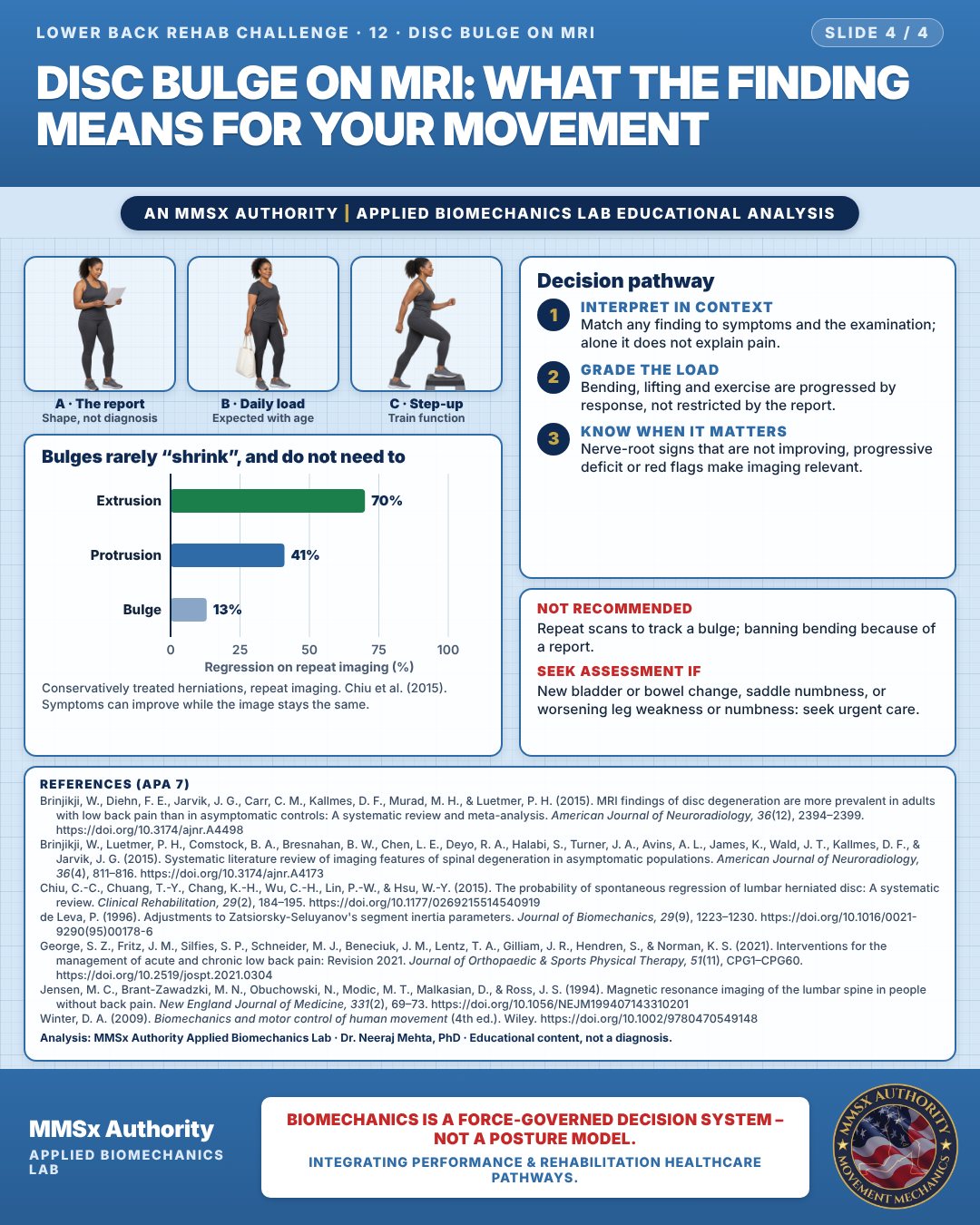
Bulges rarely “shrink”, and do not need to
Interpret in context
Match any finding to symptoms and the examination; alone it does not explain pain.
Grade the load
Bending, lifting and exercise are progressed by response, not restricted by the report.
Know when it matters
Nerve-root signs that are not improving, progressive deficit or red flags make imaging relevant.
What the evidence supports
- Interpret any MRI bulge alongside history and clinical examination; in isolation it does not identify the pain source (high prevalence in asymptomatic people, 12-A/12-B) (Brinjikji, Luetmer, et al., 2015; Jensen et al., 1994).
- Movement and exercise are managed by symptoms and function, not by the image (clinical practice guideline framework) (George et al., 2021).
- Bulges are the herniation type least likely to regress on repeat imaging (13%) (Chiu et al., 2015) — so improvement in symptoms should not be tied to a "shrinking" image; repeat scanning to track a bulge is not useful for most people.
Cautions
Not recommended
Repeat scans to track a bulge; banning bending because of a report.
Seek assessment if
New bladder or bowel change, saddle numbness, or worsening leg weakness or numbness: seek urgent care.
Methods note
2-D estimates from pose landmarks on an illustrated figure; CoM from de Leva (1996) segment parameters; cm for a 1.70 m adult (Winter, 2009). Pelvic tilt from left and right hip points in the front view. Low-back lever: upper-body CoM to an L5/S1 estimate; moment = upper-body weight × lever for a 70 kg adult, static, no hand load. These are educational estimates, not a diagnosis.
References
- Brinjikji, W., Diehn, F. E., Jarvik, J. G., Carr, C. M., Kallmes, D. F., Murad, M. H., & Luetmer, P. H. (2015). MRI findings of disc degeneration are more prevalent in adults with low back pain than in asymptomatic controls: A systematic review and meta-analysis. American Journal of Neuroradiology, 36(12), 2394–2399. https://doi.org/10.3174/ajnr.A4498
- Brinjikji, W., Luetmer, P. H., Comstock, B. A., Bresnahan, B. W., Chen, L. E., Deyo, R. A., Halabi, S., Turner, J. A., Avins, A. L., James, K., Wald, J. T., Kallmes, D. F., & Jarvik, J. G. (2015). Systematic literature review of imaging features of spinal degeneration in asymptomatic populations. American Journal of Neuroradiology, 36(4), 811–816. https://doi.org/10.3174/ajnr.A4173
- Chiu, C.-C., Chuang, T.-Y., Chang, K.-H., Wu, C.-H., Lin, P.-W., & Hsu, W.-Y. (2015). The probability of spontaneous regression of lumbar herniated disc: A systematic review. Clinical Rehabilitation, 29(2), 184–195. https://doi.org/10.1177/0269215514540919
- de Leva, P. (1996). Adjustments to Zatsiorsky-Seluyanov's segment inertia parameters. Journal of Biomechanics, 29(9), 1223–1230. https://doi.org/10.1016/0021-9290(95)00178-6
- George, S. Z., Fritz, J. M., Silfies, S. P., Schneider, M. J., Beneciuk, J. M., Lentz, T. A., Gilliam, J. R., Hendren, S., & Norman, K. S. (2021). Interventions for the management of acute and chronic low back pain: Revision 2021. Journal of Orthopaedic & Sports Physical Therapy, 51(11), CPG1–CPG60. https://doi.org/10.2519/jospt.2021.0304
- Jensen, M. C., Brant-Zawadzki, M. N., Obuchowski, N., Modic, M. T., Malkasian, D., & Ross, J. S. (1994). Magnetic resonance imaging of the lumbar spine in people without back pain. New England Journal of Medicine, 331(2), 69–73. https://doi.org/10.1056/NEJM199407143310201
- Winter, D. A. (2009). Biomechanics and motor control of human movement (4th ed.). Wiley. https://doi.org/10.1002/9780470549148
Cite this article
Mehta, N. (2026, October 23). Disc bulge on MRI: what the finding means for your movement. MMSx Authority Applied Biomechanics Lab. https://mmsxauthority.org/edu/analysis/lower-back/12-disc-bulge-on-mri-what-the-finding-means-for-your-movement/
Part of the collection
Mehta, N. (2026). Lower Back Rehabilitation: 50 Evidence-Based Biomechanics Analyses [Collection]. MMSx Authority Applied Biomechanics Lab. Zenodo. https://doi.org/10.5281/zenodo.23735778
All Lower Back Rehab Challenge articles · Rehabilitation Library