Lower Back Rehab Challenge · 10
Do you need an MRI for lower back pain? When imaging changes care
Selective imaging means requesting MRI or other imaging only when the result is likely to change management — chiefly when serious pathology is…
Key points
- Examine first. History, neurological examination and function decide whether imaging could change care.
- Image selectively. Red flags, progressive deficit, or persistent radicular pain before injection or surgery.
- Interpret in context. Read findings against age-matched pain-free people and against the examination.
What it is
Selective imaging means requesting MRI or other imaging only when the result is likely to change management — chiefly when serious pathology is suspected (e.g., fracture, malignancy, infection, cauda equina syndrome) or when persistent radicular symptoms make a person a candidate for injection or surgery — and interpreting any finding in the light of age-related prevalence, symptoms and the clinical examination.
How we analysed the movement
Three movements relevant to this topic were analysed from illustrated reference figures. Joint centres were located with two-dimensional pose estimation; joint angles, the whole-body centre of mass and, where relevant, the low-back lever (the horizontal distance from the upper-body centre of mass to the L5/S1 joint) were then calculated from standard segment parameters. The figures are educational estimates that show how position changes mechanical demand; they are not measurements of a patient. Every research value quoted below comes from the published source cited beside it.
Movement A: Questions about a scan (front view)
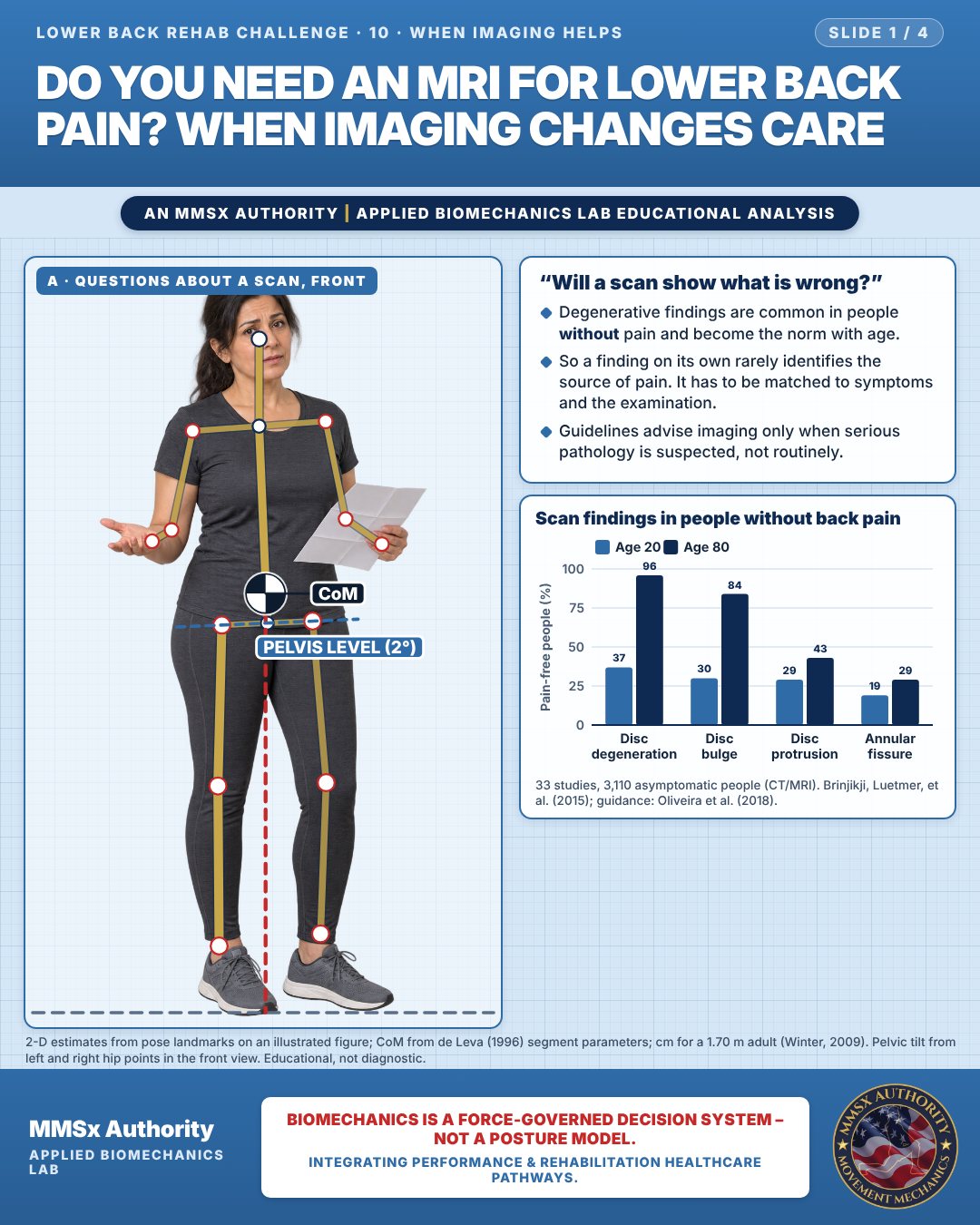
“Will a scan show what is wrong?”
Degenerative findings are common in people without pain and become the norm with age.
So a finding on its own rarely identifies the source of pain. It has to be matched to symptoms and the examination.
Guidelines advise imaging only when serious pathology is suspected, not routinely.
Scan findings in people without back pain
Movement B: Functional testing (side view)
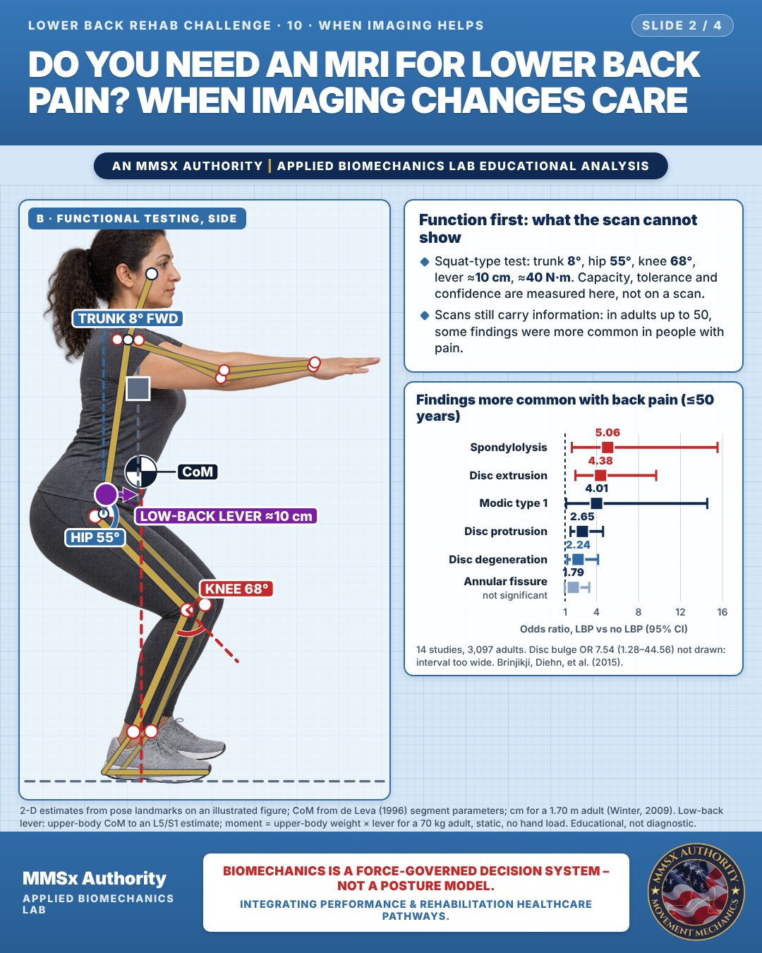
Function first: what the scan cannot show
Squat-type test: trunk 8°, hip 55°, knee 68°, lever ≈10 cm, ≈40 N·m. Capacity, tolerance and confidence are measured here, not on a scan.
Scans still carry information: in adults up to 50, some findings were more common in people with pain.
Findings more common with back pain (≤50 years)
Movement C: Discussing the plan
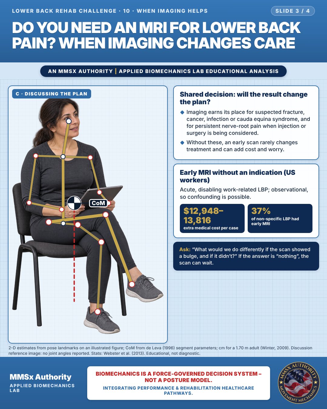
Shared decision: will the result change the plan?
Imaging earns its place for suspected fracture, cancer, infection or cauda equina syndrome, and for persistent nerve-root pain when injection or surgery is being considered.
Without these, an early scan rarely changes treatment and can add cost and worry.
Early MRI without an indication (US workers)
Acute, disabling work-related LBP; observational, so confounding is possible.
Decision pathway
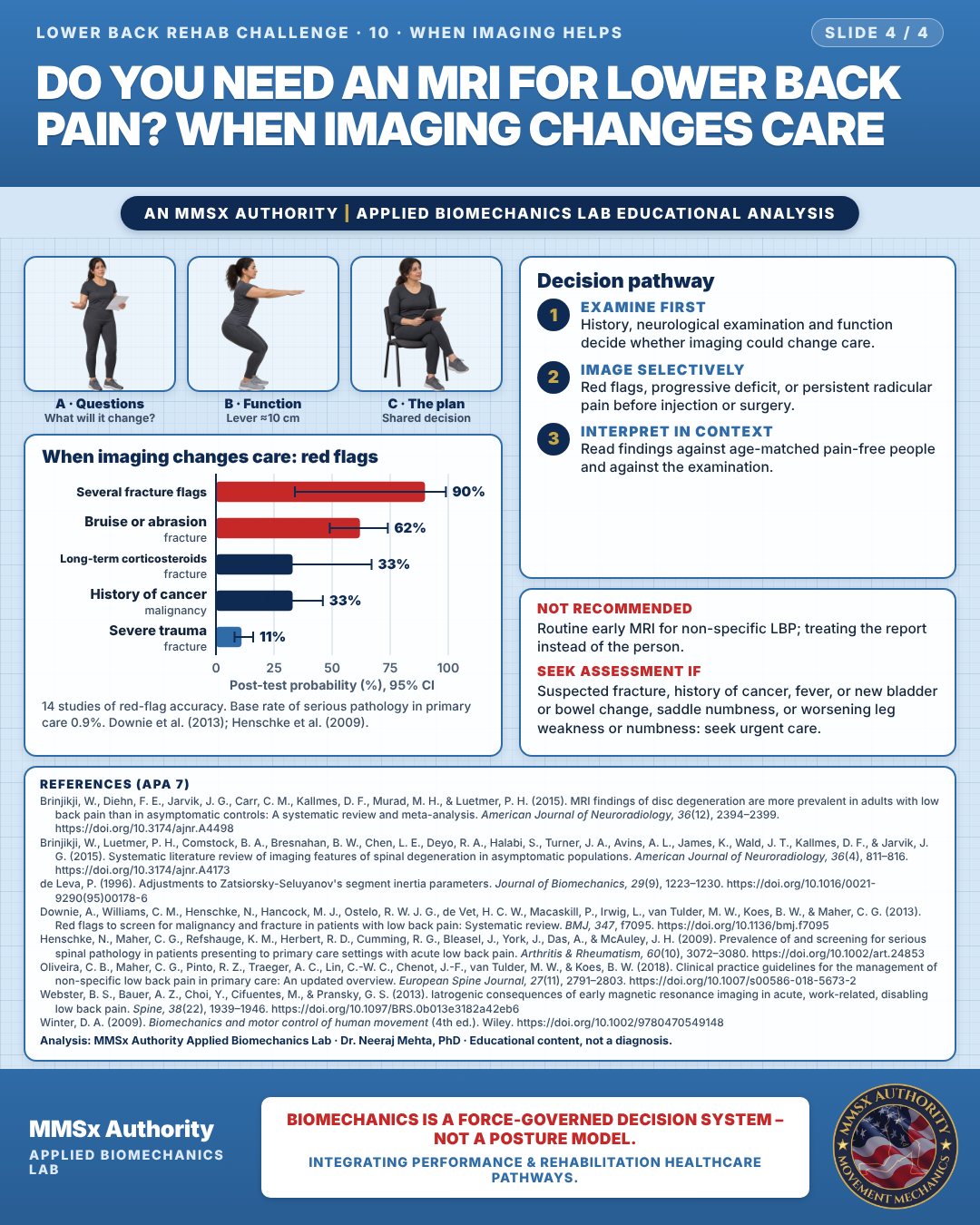
When imaging changes care: red flags
Examine first
History, neurological examination and function decide whether imaging could change care.
Image selectively
Red flags, progressive deficit, or persistent radicular pain before injection or surgery.
Interpret in context
Read findings against age-matched pain-free people and against the examination.
What the evidence supports
- Imaging only if serious pathology is suspected; routine imaging NOT recommended. (Oliveira et al., 2018)
- MRI is generally reserved for persistent radicular pain after about 1 month of conservative care in people who are candidates for surgery or epidural injection, or for red flags (as summarised in the background of Webster et al., 2013).
- When imaging is done, interpret findings against age-specific prevalence in people without pain and against the examination. (Brinjikji et al., 2015b, 2015a)
Myth and evidence
Myth“An MRI will find the cause of my pain.”
EvidenceDisc degeneration and bulges are present in most pain-free older adults (96% and 84% at 80). (Brinjikji et al., 2015b)
Myth“Scans are useless.”
EvidenceSeveral findings (e.g., extrusion, Modic 1, spondylolysis) are more common in younger adults with pain, and imaging is essential when red flags or surgical decisions are present. (Brinjikji et al., 2015a; Downie et al., 2013)
Cautions
Not recommended
Routine early MRI for non-specific LBP; treating the report instead of the person.
Seek assessment if
Suspected fracture, history of cancer, fever, or new bladder or bowel change, saddle numbness, or worsening leg weakness or numbness: seek urgent care.
Methods note
2-D estimates from pose landmarks on an illustrated figure; CoM from de Leva (1996) segment parameters; cm for a 1.70 m adult (Winter, 2009). Pelvic tilt from left and right hip points in the front view. Low-back lever: upper-body CoM to an L5/S1 estimate; moment = upper-body weight × lever for a 70 kg adult, static, no hand load. Discussion reference image: no joint angles reported. Stats: Webster et al. (2013). These are educational estimates, not a diagnosis.
References
- Brinjikji, W., Diehn, F. E., Jarvik, J. G., Carr, C. M., Kallmes, D. F., Murad, M. H., & Luetmer, P. H. (2015). MRI findings of disc degeneration are more prevalent in adults with low back pain than in asymptomatic controls: A systematic review and meta-analysis. American Journal of Neuroradiology, 36(12), 2394–2399. https://doi.org/10.3174/ajnr.A4498
- Brinjikji, W., Luetmer, P. H., Comstock, B. A., Bresnahan, B. W., Chen, L. E., Deyo, R. A., Halabi, S., Turner, J. A., Avins, A. L., James, K., Wald, J. T., Kallmes, D. F., & Jarvik, J. G. (2015). Systematic literature review of imaging features of spinal degeneration in asymptomatic populations. American Journal of Neuroradiology, 36(4), 811–816. https://doi.org/10.3174/ajnr.A4173
- de Leva, P. (1996). Adjustments to Zatsiorsky-Seluyanov's segment inertia parameters. Journal of Biomechanics, 29(9), 1223–1230. https://doi.org/10.1016/0021-9290(95)00178-6
- Downie, A., Williams, C. M., Henschke, N., Hancock, M. J., Ostelo, R. W. J. G., de Vet, H. C. W., Macaskill, P., Irwig, L., van Tulder, M. W., Koes, B. W., & Maher, C. G. (2013). Red flags to screen for malignancy and fracture in patients with low back pain: Systematic review. BMJ, 347, f7095. https://doi.org/10.1136/bmj.f7095
- Henschke, N., Maher, C. G., Refshauge, K. M., Herbert, R. D., Cumming, R. G., Bleasel, J., York, J., Das, A., & McAuley, J. H. (2009). Prevalence of and screening for serious spinal pathology in patients presenting to primary care settings with acute low back pain. Arthritis & Rheumatism, 60(10), 3072–3080. https://doi.org/10.1002/art.24853
- Oliveira, C. B., Maher, C. G., Pinto, R. Z., Traeger, A. C., Lin, C.-W. C., Chenot, J.-F., van Tulder, M. W., & Koes, B. W. (2018). Clinical practice guidelines for the management of non-specific low back pain in primary care: An updated overview. European Spine Journal, 27(11), 2791–2803. https://doi.org/10.1007/s00586-018-5673-2
- Webster, B. S., Bauer, A. Z., Choi, Y., Cifuentes, M., & Pransky, G. S. (2013). Iatrogenic consequences of early magnetic resonance imaging in acute, work-related, disabling low back pain. Spine, 38(22), 1939–1946. https://doi.org/10.1097/BRS.0b013e3182a42eb6
- Winter, D. A. (2009). Biomechanics and motor control of human movement (4th ed.). Wiley. https://doi.org/10.1002/9780470549148
Cite this article
Mehta, N. (2026, October 23). Do you need an MRI for lower back pain? When imaging changes care. MMSx Authority Applied Biomechanics Lab. https://mmsxauthority.org/edu/analysis/lower-back/10-do-you-need-an-mri-for-lower-back-pain-when-imaging-changes-care/
Part of the collection
Mehta, N. (2026). Lower Back Rehabilitation: 50 Evidence-Based Biomechanics Analyses [Collection]. MMSx Authority Applied Biomechanics Lab. Zenodo. https://doi.org/10.5281/zenodo.23735778
All Lower Back Rehab Challenge articles · Rehabilitation Library