Knee Rehab Challenge · 44
When knee pain comes from the hip
Referred knee pain from the hip is pain felt in the thigh or knee that originates from hip joint pathology (e.g.
Key points
- Examine the hip. In every knee-pain assessment: hip rotation (especially internal rotation) and pain provocation.
- Listen for clues. Groin pain (sensitivity 84%) or knee pain with an unremarkable knee examination points to the hip.
- Act fast in teens. Knee or thigh pain with a limp in a young person: same-day assessment for SCFE.
What it is
Referred knee pain from the hip is pain felt in the thigh or knee that originates from hip joint pathology (e.g. hip osteoarthritis, osteonecrosis, or — in adolescents — slipped capital femoral epiphysis, SCFE). The knee itself may examine normally, so the hip should be examined in anyone with knee pain whose knee findings do not explain the symptoms.
How we analysed the movement
Three movements relevant to this topic were analysed from illustrated reference figures. Joint centres were located with two-dimensional pose estimation; joint angles, the whole-body centre of mass and, where relevant, the ground-reaction-force line and its moment arm about the knee were then calculated from standard segment parameters. The figures are educational estimates that show how position changes mechanical demand; they are not measurements of a patient. Every research value quoted below comes from the published source cited beside it.
Movement A: Walking (side view)
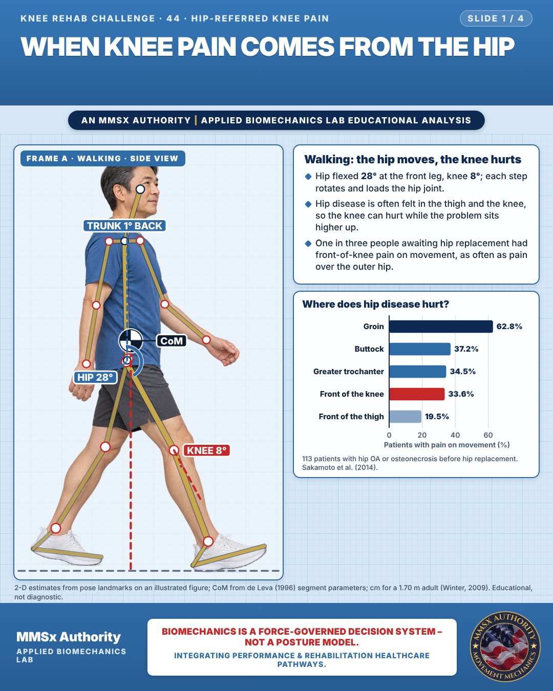
Walking: the hip moves, the knee hurts
Hip flexed 28° at the front leg, knee 8°; each step rotates and loads the hip joint.
Hip disease is often felt in the thigh and the knee, so the knee can hurt while the problem sits higher up.
One in three people awaiting hip replacement had front-of-knee pain on movement, as often as pain over the outer hip.
Where does hip disease hurt?
Movement B: Walking (front view)
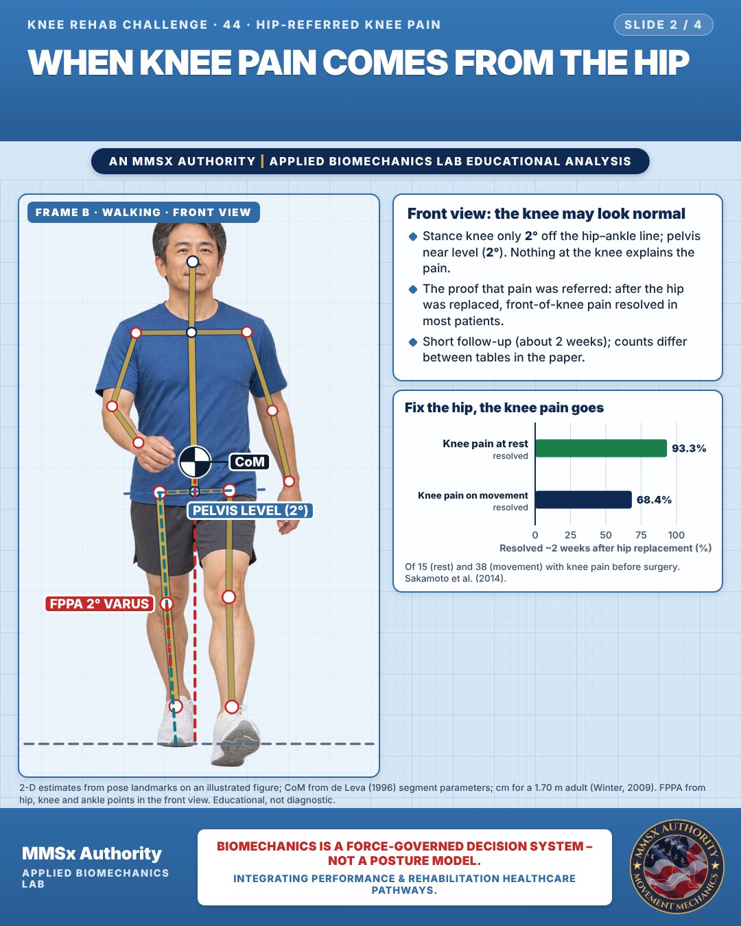
Front view: the knee may look normal
Stance knee only 2° off the hip–ankle line; pelvis near level (2°). Nothing at the knee explains the pain.
The proof that pain was referred: after the hip was replaced, front-of-knee pain resolved in most patients.
Short follow-up (about 2 weeks); counts differ between tables in the paper.
Fix the hip, the knee pain goes
Movement C: Walking (rear view)
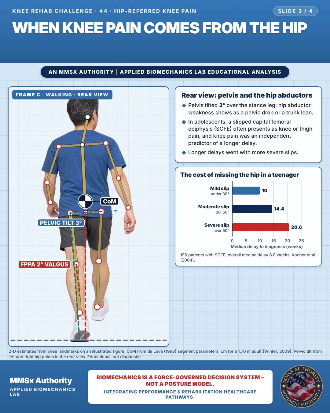
Rear view: pelvis and the hip abductors
Pelvis tilted 3° over the stance leg; hip abductor weakness shows as a pelvic drop or a trunk lean.
In adolescents, a slipped capital femoral epiphysis (SCFE) often presents as knee or thigh pain, and knee pain was an independent predictor of a longer delay.
Longer delays went with more severe slips.
The cost of missing the hip in a teenager
Decision pathway
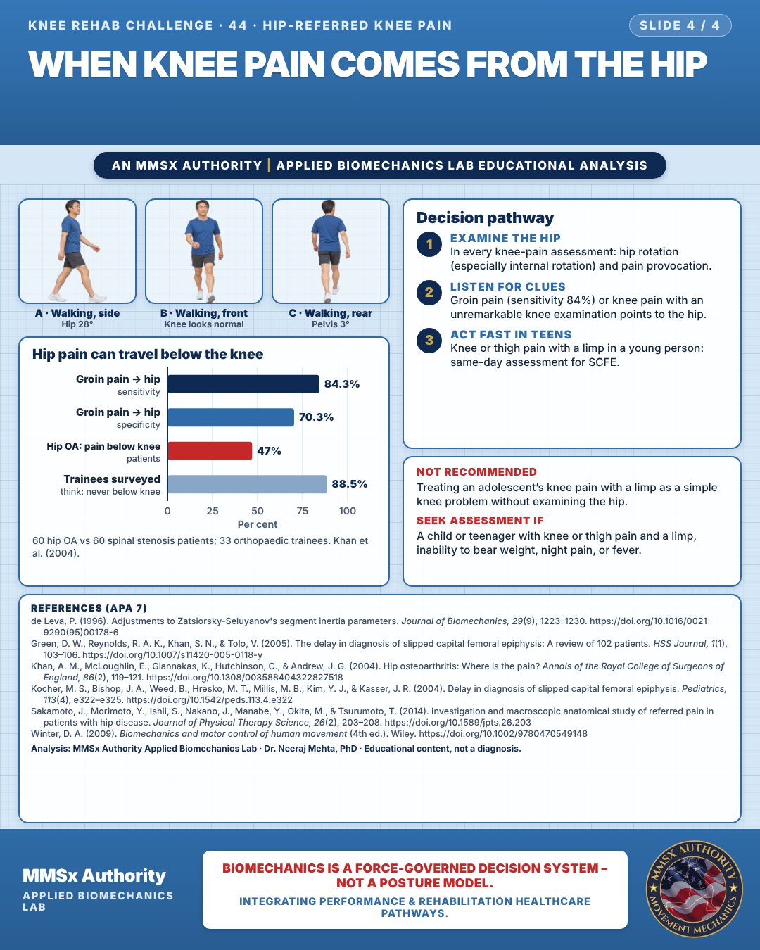
Hip pain can travel below the knee
Examine the hip
In every knee-pain assessment: hip rotation (especially internal rotation) and pain provocation.
Listen for clues
Groin pain (sensitivity 84%) or knee pain with an unremarkable knee examination points to the hip.
Act fast in teens
Knee or thigh pain with a limp in a young person: same-day assessment for SCFE.
What the evidence supports
- Recommend: examine the hip (range of motion, especially internal rotation, and pain provocation) in every patient presenting with knee or thigh pain, particularly when knee examination is unremarkable (Khan 2004; Sakamoto 2014; Kocher 2004 conclusion — emphasis on knee/thigh-pain presentations).
Myth and evidence
Myth“Hip pain never goes below the knee.”
Evidence47% of hip OA patients had pain below the knee while 88.5% of surveyed trainees held the opposite belief (Khan 2004); one-third of hip patients reported anterior knee pain on movement, which in most resolved after hip replacement (Sakamoto 2014).
Cautions
Not recommended
Treating an adolescent’s knee pain with a limp as a simple knee problem without examining the hip.
Seek assessment if
A child or teenager with knee or thigh pain and a limp, inability to bear weight, night pain, or fever.
Methods note
2-D estimates from pose landmarks on an illustrated figure; CoM from de Leva (1996) segment parameters; cm for a 1.70 m adult (Winter, 2009). FPPA from hip, knee and ankle points in the front view. Pelvic tilt from left and right hip points in the rear view. These are educational estimates, not a diagnosis.
References
- de Leva, P. (1996). Adjustments to Zatsiorsky-Seluyanov's segment inertia parameters. Journal of Biomechanics, 29(9), 1223–1230. https://doi.org/10.1016/0021-9290(95)00178-6
- Green, D. W., Reynolds, R. A. K., Khan, S. N., & Tolo, V. (2005). The delay in diagnosis of slipped capital femoral epiphysis: A review of 102 patients. HSS Journal, 1(1), 103–106. https://doi.org/10.1007/s11420-005-0118-y
- Khan, A. M., McLoughlin, E., Giannakas, K., Hutchinson, C., & Andrew, J. G. (2004). Hip osteoarthritis: Where is the pain? Annals of the Royal College of Surgeons of England, 86(2), 119–121. https://doi.org/10.1308/003588404322827518
- Kocher, M. S., Bishop, J. A., Weed, B., Hresko, M. T., Millis, M. B., Kim, Y. J., & Kasser, J. R. (2004). Delay in diagnosis of slipped capital femoral epiphysis. Pediatrics, 113(4), e322–e325. https://doi.org/10.1542/peds.113.4.e322
- Sakamoto, J., Morimoto, Y., Ishii, S., Nakano, J., Manabe, Y., Okita, M., & Tsurumoto, T. (2014). Investigation and macroscopic anatomical study of referred pain in patients with hip disease. Journal of Physical Therapy Science, 26(2), 203–208. https://doi.org/10.1589/jpts.26.203
- Winter, D. A. (2009). Biomechanics and motor control of human movement (4th ed.). Wiley. https://doi.org/10.1002/9780470549148
Cite this article
Mehta, N. (2026, October 23). When knee pain comes from the hip. MMSx Authority Applied Biomechanics Lab. https://mmsxauthority.org/edu/analysis/knee/44-when-knee-pain-comes-from-the-hip/
Part of the collection
Mehta, N. (2026). Knee Rehabilitation: 50 Evidence-Based Biomechanics Analyses [Collection]. MMSx Authority Applied Biomechanics Lab. Zenodo. https://doi.org/10.5281/zenodo.23735752