Knee Rehab Challenge · 42
Knee pain in pregnancy: load, posture and safe movement
Knee pain in pregnancy refers to new or worsened knee symptoms during pregnancy. Movement analysis, verified research data and a decision pathway from the MMS
Key points
- Keep moving. Pregnant women should be encouraged to continue or begin safe physical activity (ACOG, 2020).
- Share the load. Favour two-legged, supported squats and use the handrail on stairs; hold loads close.
- Strengthen hips. Hip and ankle muscles work harder late in pregnancy: include hip and calf strengthening.
What it is
Knee pain in pregnancy refers to new or worsened knee symptoms during pregnancy. Body mass increases, the centre of mass moves forward, and walking and stair mechanics adapt, which changes how the knees are loaded; hormonal changes may also influence soft tissues. Most knee symptoms in pregnancy are load-related and respond to activity adjustment and exercise, but swelling of one calf or leg needs urgent medical review.
How we analysed the movement
Three movements relevant to this topic were analysed from illustrated reference figures. Joint centres were located with two-dimensional pose estimation; joint angles, the whole-body centre of mass and, where relevant, the ground-reaction-force line and its moment arm about the knee were then calculated from standard segment parameters. The figures are educational estimates that show how position changes mechanical demand; they are not measurements of a patient. Every research value quoted below comes from the published source cited beside it.
Movement A: Squat (side view)
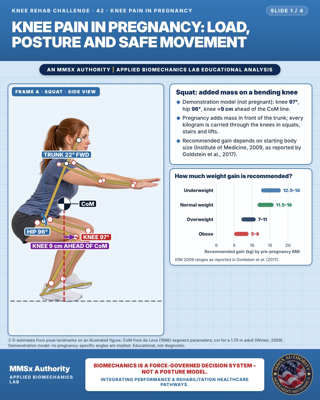
Squat: added mass on a bending knee
Demonstration model (not pregnant): knee 97°, hip 96°, knee ≈9 cm ahead of the CoM line.
Pregnancy adds mass in front of the trunk; every kilogram is carried through the knees in squats, stairs and lifts.
Recommended gain depends on starting body size (Institute of Medicine, 2009, as reported by Goldstein et al., 2017).
How much weight gain is recommended?
Movement B: Squat (front view)
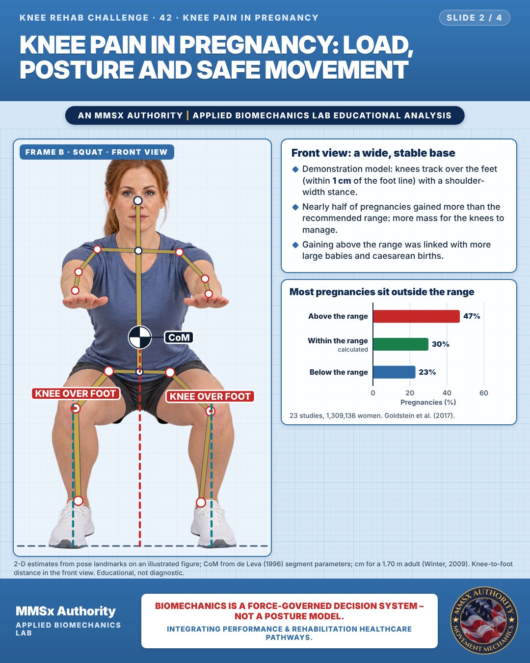
Front view: a wide, stable base
Demonstration model: knees track over the feet (within 1 cm of the foot line) with a shoulder-width stance.
Nearly half of pregnancies gained more than the recommended range: more mass for the knees to manage.
Gaining above the range was linked with more large babies and caesarean births.
Most pregnancies sit outside the range
Movement C: Lifting a box (side view)
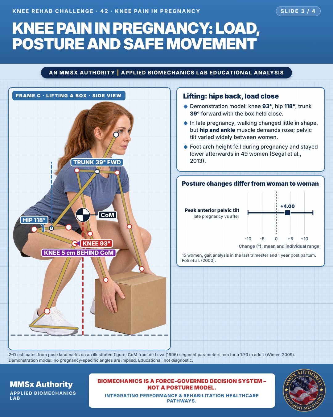
Lifting: hips back, load close
Demonstration model: knee 93°, hip 118°, trunk 39° forward with the box held close.
In late pregnancy, walking changed little in shape, but hip and ankle muscle demands rose; pelvic tilt varied widely between women.
Foot arch height fell during pregnancy and stayed lower afterwards in 49 women (Segal et al., 2013).
Posture changes differ from woman to woman
Decision pathway
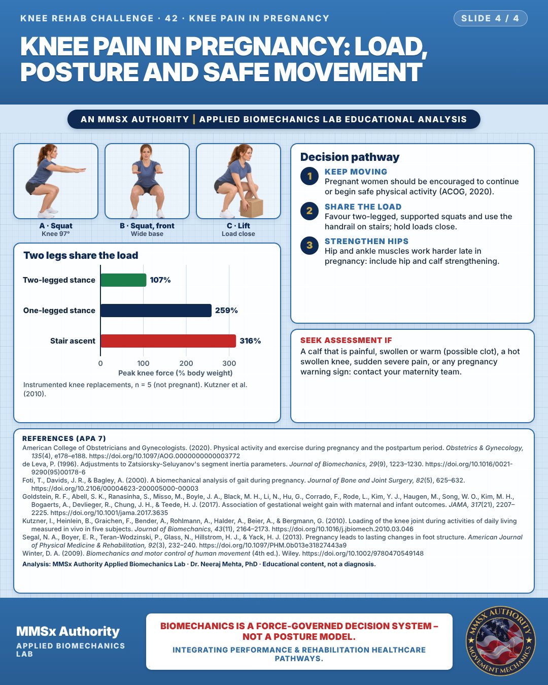
Two legs share the load
Keep moving
Pregnant women should be encouraged to continue or begin safe physical activity (ACOG, 2020).
Share the load
Favour two-legged, supported squats and use the handrail on stairs; hold loads close.
Strengthen hips
Hip and ankle muscles work harder late in pregnancy: include hip and calf strengthening.
Cautions
Seek assessment if
A calf that is painful, swollen or warm (possible clot), a hot swollen knee, sudden severe pain, or any pregnancy warning sign: contact your maternity team.
Methods note
2-D estimates from pose landmarks on an illustrated figure; CoM from de Leva (1996) segment parameters; cm for a 1.70 m adult (Winter, 2009). Demonstration model: no pregnancy-specific angles are implied. Knee-to-foot distance in the front view. These are educational estimates, not a diagnosis.
References
- American College of Obstetricians and Gynecologists. (2020). Physical activity and exercise during pregnancy and the postpartum period. Obstetrics & Gynecology, 135(4), e178–e188. https://doi.org/10.1097/AOG.0000000000003772
- de Leva, P. (1996). Adjustments to Zatsiorsky-Seluyanov's segment inertia parameters. Journal of Biomechanics, 29(9), 1223–1230. https://doi.org/10.1016/0021-9290(95)00178-6
- Foti, T., Davids, J. R., & Bagley, A. (2000). A biomechanical analysis of gait during pregnancy. Journal of Bone and Joint Surgery, 82(5), 625–632. https://doi.org/10.2106/00004623-200005000-00003
- Goldstein, R. F., Abell, S. K., Ranasinha, S., Misso, M., Boyle, J. A., Black, M. H., Li, N., Hu, G., Corrado, F., Rode, L., Kim, Y. J., Haugen, M., Song, W. O., Kim, M. H., Bogaerts, A., Devlieger, R., Chung, J. H., & Teede, H. J. (2017). Association of gestational weight gain with maternal and infant outcomes. JAMA, 317(21), 2207–2225. https://doi.org/10.1001/jama.2017.3635
- Kutzner, I., Heinlein, B., Graichen, F., Bender, A., Rohlmann, A., Halder, A., Beier, A., & Bergmann, G. (2010). Loading of the knee joint during activities of daily living measured in vivo in five subjects. Journal of Biomechanics, 43(11), 2164–2173. https://doi.org/10.1016/j.jbiomech.2010.03.046
- Segal, N. A., Boyer, E. R., Teran-Wodzinski, P., Glass, N., Hillstrom, H. J., & Yack, H. J. (2013). Pregnancy leads to lasting changes in foot structure. American Journal of Physical Medicine & Rehabilitation, 92(3), 232–240. https://doi.org/10.1097/PHM.0b013e31827443a9
- Winter, D. A. (2009). Biomechanics and motor control of human movement (4th ed.). Wiley. https://doi.org/10.1002/9780470549148
Cite this article
Mehta, N. (2026, October 23). Knee pain in pregnancy: load, posture and safe movement. MMSx Authority Applied Biomechanics Lab. https://mmsxauthority.org/edu/analysis/knee/42-knee-pain-in-pregnancy-load-posture-and-safe-movement/
Part of the collection
Mehta, N. (2026). Knee Rehabilitation: 50 Evidence-Based Biomechanics Analyses [Collection]. MMSx Authority Applied Biomechanics Lab. Zenodo. https://doi.org/10.5281/zenodo.23735752