Knee Rehab Challenge · 40
Plica syndrome: a painful fold inside the knee
A plica is a normal fold of the synovial lining inside the knee joint. Movement analysis, verified research data and a decision pathway from the MMSx Authorit
Key points
- Rule out others. A plica alone was the finding in only about 4% of arthroscopies: rule out commoner causes of front-inner knee pain first.
- Treat conservatively. Flexibility of the hamstrings, calf and quadriceps; reduce repeated deep bending for a while; strengthen hip and knee.
- Refer if persistent. Surgery suited selected patients with a specific diagnosis and impingement findings.
What it is
A plica is a normal fold of the synovial lining inside the knee joint; the medial plica is the most commonly described. Plica syndrome refers to pain, and sometimes clicking or catching, when a plica becomes thickened or irritated and is pinched during knee bending and straightening. Because plicae are common in knees without symptoms, the diagnosis is made by excluding other causes.
How we analysed the movement
Three movements relevant to this topic were analysed from illustrated reference figures. Joint centres were located with two-dimensional pose estimation; joint angles, the whole-body centre of mass and, where relevant, the ground-reaction-force line and its moment arm about the knee were then calculated from standard segment parameters. The figures are educational estimates that show how position changes mechanical demand; they are not measurements of a patient. Every research value quoted below comes from the published source cited beside it.
Movement A: Pedal near the top (side view)
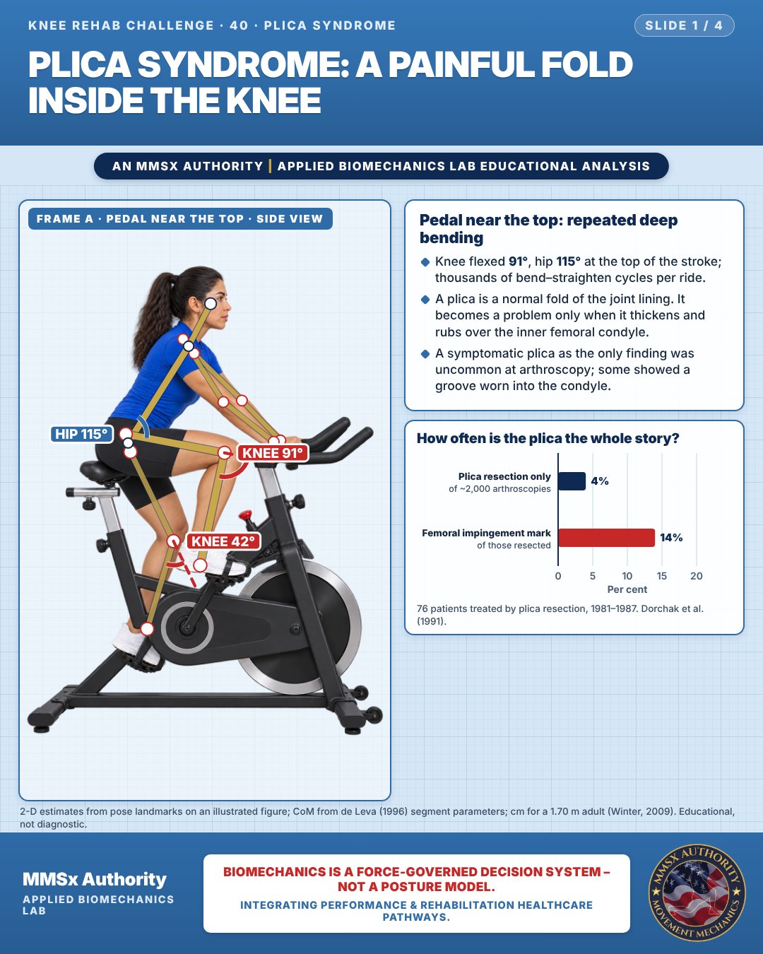
Pedal near the top: repeated deep bending
Knee flexed 91°, hip 115° at the top of the stroke; thousands of bend–straighten cycles per ride.
A plica is a normal fold of the joint lining. It becomes a problem only when it thickens and rubs over the inner femoral condyle.
A symptomatic plica as the only finding was uncommon at arthroscopy; some showed a groove worn into the condyle.
How often is the plica the whole story?
Movement B: Step-down (front view)
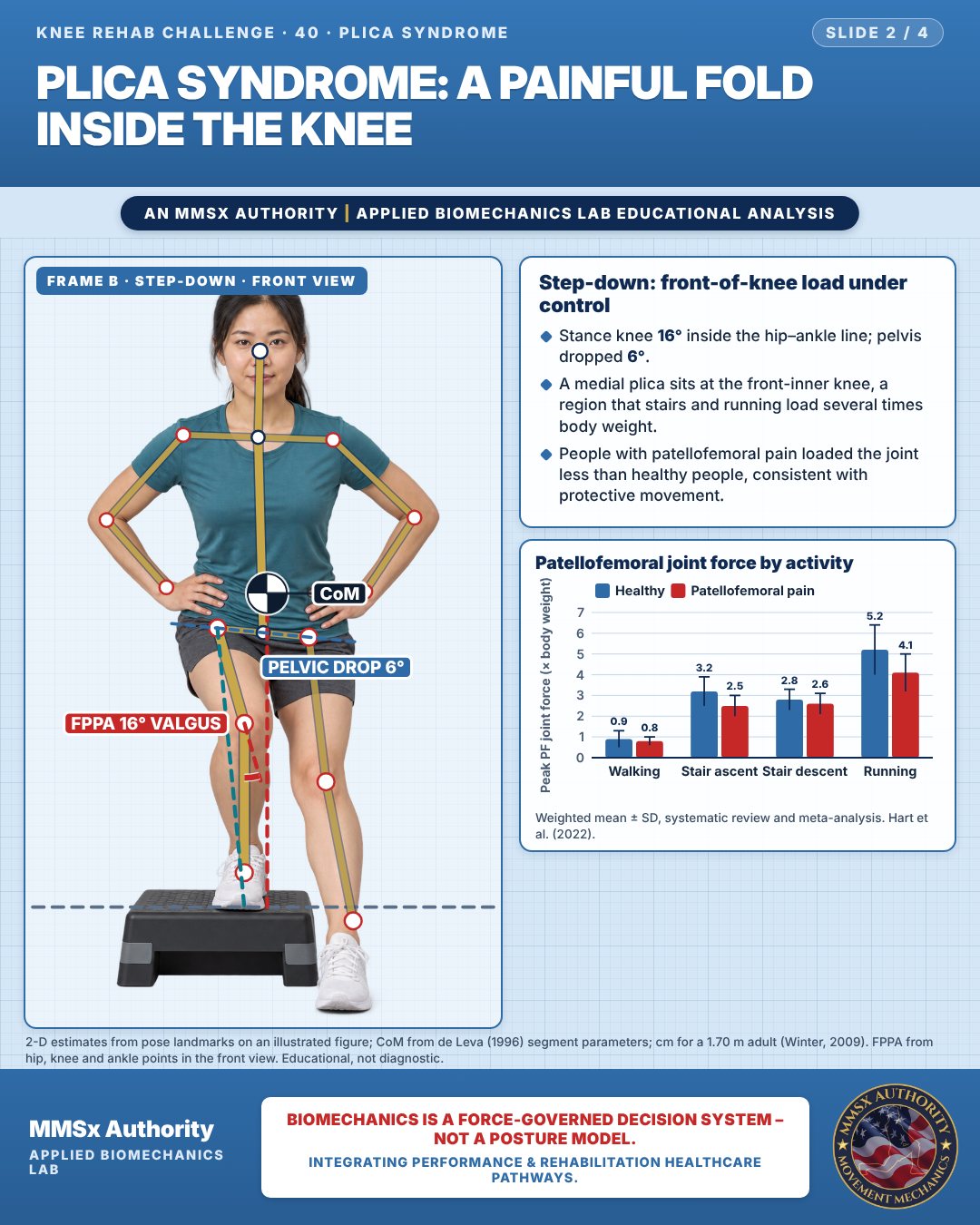
Step-down: front-of-knee load under control
Stance knee 16° inside the hip–ankle line; pelvis dropped 6°.
A medial plica sits at the front-inner knee, a region that stairs and running load several times body weight.
People with patellofemoral pain loaded the joint less than healthy people, consistent with protective movement.
Patellofemoral joint force by activity
Movement C: Seated, knee bent (side view)
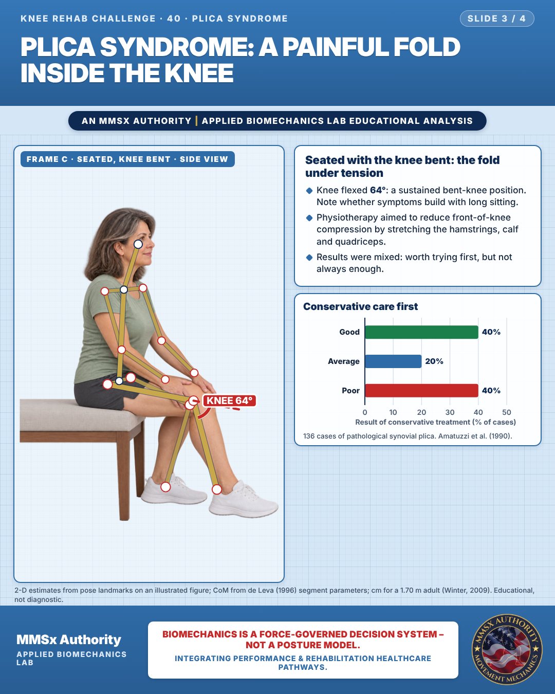
Seated with the knee bent: the fold under tension
Knee flexed 64°: a sustained bent-knee position. Note whether symptoms build with long sitting.
Physiotherapy aimed to reduce front-of-knee compression by stretching the hamstrings, calf and quadriceps.
Results were mixed: worth trying first, but not always enough.
Conservative care first
Decision pathway
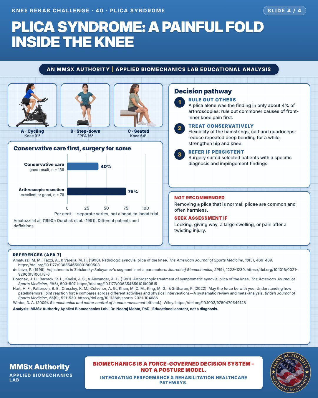
Conservative care first, surgery for some
Rule out others
A plica alone was the finding in only about 4% of arthroscopies: rule out commoner causes of front-inner knee pain first.
Treat conservatively
Flexibility of the hamstrings, calf and quadriceps; reduce repeated deep bending for a while; strengthen hip and knee.
Refer if persistent
Surgery suited selected patients with a specific diagnosis and impingement findings.
Cautions
Not recommended
Removing a plica that is normal: plicae are common and often harmless.
Seek assessment if
Locking, giving way, a large swelling, or pain after a twisting injury.
Methods note
2-D estimates from pose landmarks on an illustrated figure; CoM from de Leva (1996) segment parameters; cm for a 1.70 m adult (Winter, 2009). FPPA from hip, knee and ankle points in the front view. These are educational estimates, not a diagnosis.
References
- Amatuzzi, M. M., Fazzi, A., & Varella, M. H. (1990). Pathologic synovial plica of the knee. The American Journal of Sports Medicine, 18(5), 466–469. https://doi.org/10.1177/036354659001800503
- de Leva, P. (1996). Adjustments to Zatsiorsky-Seluyanov's segment inertia parameters. Journal of Biomechanics, 29(9), 1223–1230. https://doi.org/10.1016/0021-9290(95)00178-6
- Dorchak, J. D., Barrack, R. L., Kneisl, J. S., & Alexander, A. H. (1991). Arthroscopic treatment of symptomatic synovial plica of the knee. The American Journal of Sports Medicine, 19(5), 503–507. https://doi.org/10.1177/036354659101900515
- Hart, H. F., Patterson, B. E., Crossley, K. M., Culvenor, A. G., Khan, M. C. M., King, M. G., & Sritharan, P. (2022). May the force be with you: Understanding how patellofemoral joint reaction force compares across different activities and physical interventions—A systematic review and meta-analysis. British Journal of Sports Medicine, 56(9), 521–530. https://doi.org/10.1136/bjsports-2021-104686
- Winter, D. A. (2009). Biomechanics and motor control of human movement (4th ed.). Wiley. https://doi.org/10.1002/9780470549148
Cite this article
Mehta, N. (2026, October 23). Plica syndrome: a painful fold inside the knee. MMSx Authority Applied Biomechanics Lab. https://mmsxauthority.org/edu/analysis/knee/40-plica-syndrome-a-painful-fold-inside-the-knee/
Part of the collection
Mehta, N. (2026). Knee Rehabilitation: 50 Evidence-Based Biomechanics Analyses [Collection]. MMSx Authority Applied Biomechanics Lab. Zenodo. https://doi.org/10.5281/zenodo.23735752