Knee Rehab Challenge · 39
Cartilage damage: what a "worn spot" means for knee loading
An articular cartilage defect is a localised loss or damage of the smooth hyaline cartilage covering the joint surfaces (graded at arthroscopy, e.g.
Key points
- Modify. Use low-compression movement such as cycling while symptoms are high; reduce stairs and long walks briefly.
- Strengthen. Graded, knee-loading exercise: it has not been shown to damage cartilage.
- Progress. Walking, then stairs and impact as tolerated. Surgery fits few: 7% of under-40s had repair-type lesions.
What it is
An articular cartilage defect is a localised loss or damage of the smooth hyaline cartilage covering the joint surfaces (graded at arthroscopy, e.g. Outerbridge grades I–IV, from softening to full-thickness loss exposing bone); it may be focal (single "worn spot") or part of more widespread osteoarthritic change. Cartilage has no nerves, so pain usually reflects the surrounding joint (synovium, bone, effusion), and a defect on a scan does not by itself explain symptoms.
How we analysed the movement
Three movements relevant to this topic were analysed from illustrated reference figures. Joint centres were located with two-dimensional pose estimation; joint angles, the whole-body centre of mass and, where relevant, the ground-reaction-force line and its moment arm about the knee were then calculated from standard segment parameters. The figures are educational estimates that show how position changes mechanical demand; they are not measurements of a patient. Every research value quoted below comes from the published source cited beside it.
Movement A: Walking (side view)
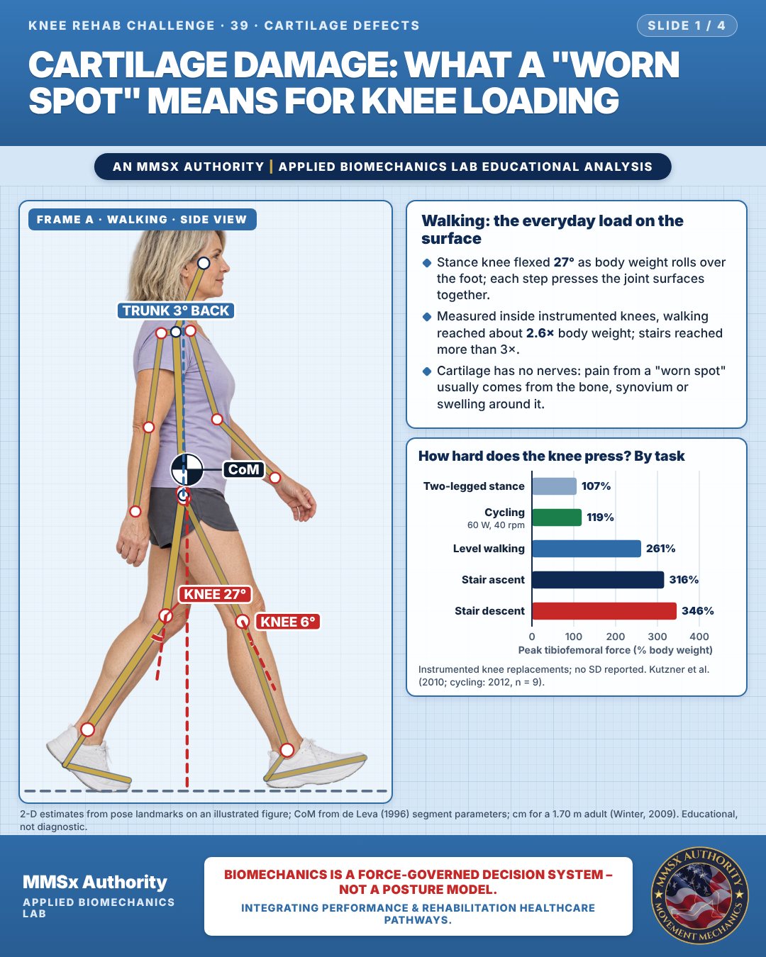
Walking: the everyday load on the surface
Stance knee flexed 27° as body weight rolls over the foot; each step presses the joint surfaces together.
Measured inside instrumented knees, walking reached about 2.6× body weight; stairs reached more than 3×.
Cartilage has no nerves: pain from a "worn spot" usually comes from the bone, synovium or swelling around it.
How hard does the knee press? By task
Movement B: Cycling, downstroke (side view)
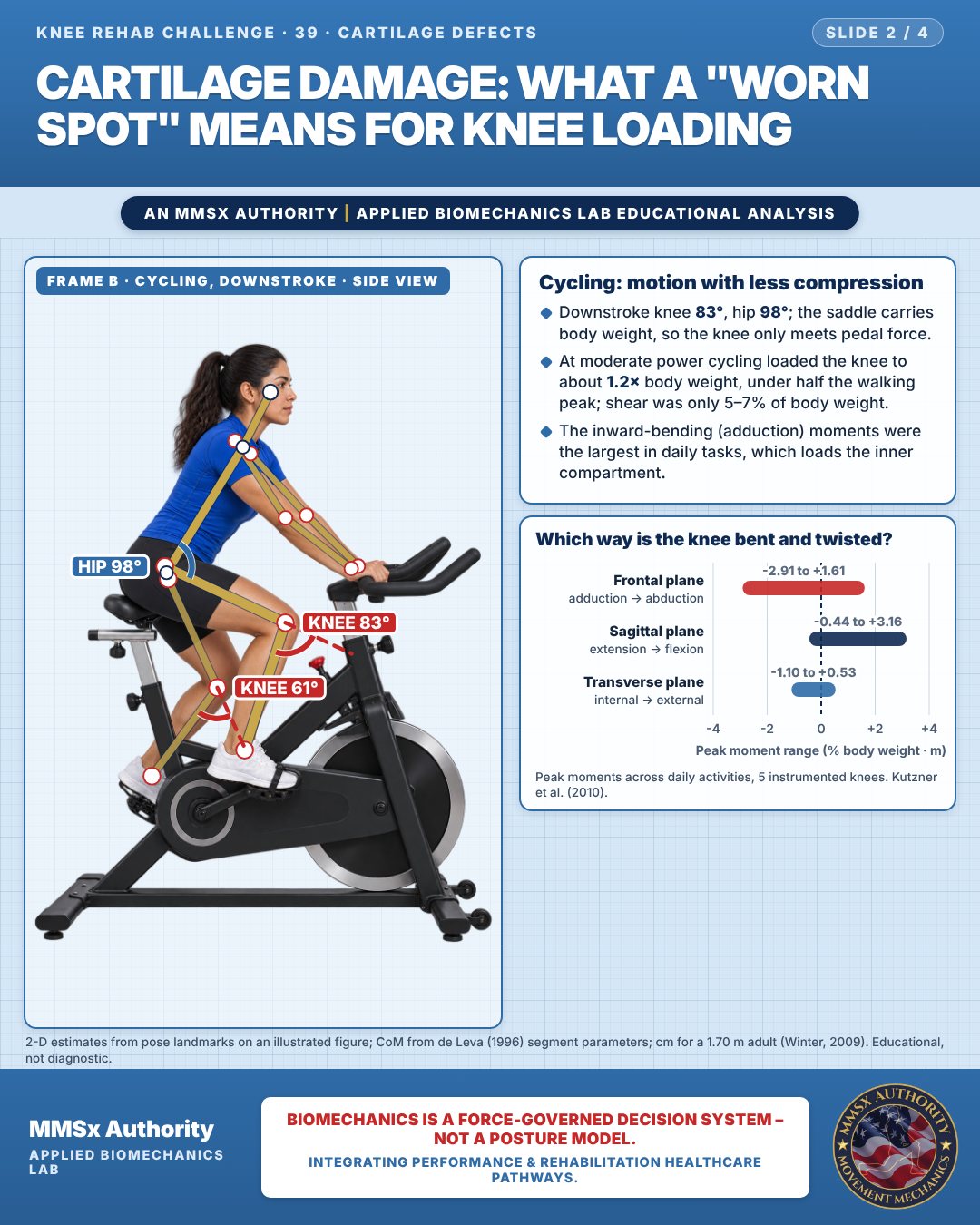
Cycling: motion with less compression
Downstroke knee 83°, hip 98°; the saddle carries body weight, so the knee only meets pedal force.
At moderate power cycling loaded the knee to about 1.2× body weight, under half the walking peak; shear was only 5–7% of body weight.
The inward-bending (adduction) moments were the largest in daily tasks, which loads the inner compartment.
Which way is the knee bent and twisted?
Movement C: Cycling (front view)
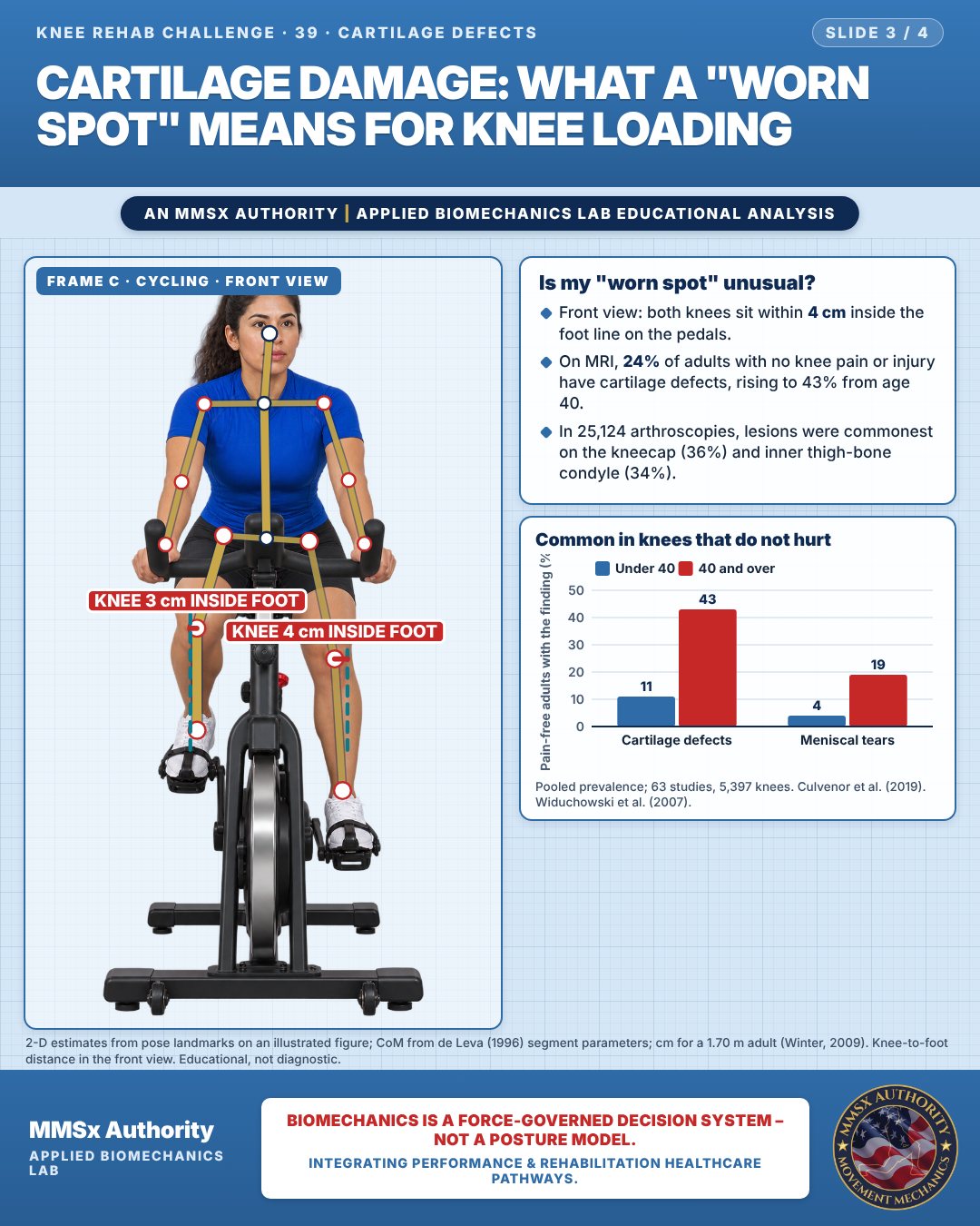
Is my "worn spot" unusual?
Front view: both knees sit within 4 cm inside the foot line on the pedals.
On MRI, 24% of adults with no knee pain or injury have cartilage defects, rising to 43% from age 40.
In 25,124 arthroscopies, lesions were commonest on the kneecap (36%) and inner thigh-bone condyle (34%).
Common in knees that do not hurt
Decision pathway
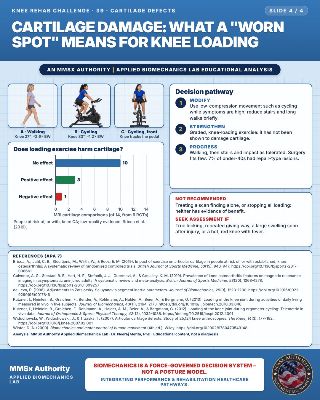
Does loading exercise harm cartilage?
Modify
Use low-compression movement such as cycling while symptoms are high; reduce stairs and long walks briefly.
Strengthen
Graded, knee-loading exercise: it has not been shown to damage cartilage.
Progress
Walking, then stairs and impact as tolerated. Surgery fits few: 7% of under-40s had repair-type lesions.
What the evidence supports
- Recommended direction: graded, knee-loading exercise is not shown to damage cartilage in people at risk of or with knee OA (Bricca et al., 2019, low-quality evidence). Low-compression options (stationary cycling ≈119% BW at moderate power vs ≈261% BW walking in implant data) let people keep moving while symptoms settle (Kutzner et al., 2010, 2012); load is then progressed (walking, stairs) as tolerated.
- Not recommended: treating an imaging finding alone — cartilage defects occur in 24% of pain-free knees (43% at ≥ 40 y) (Culvenor et al., 2019); prolonged rest/avoiding all loading (no evidence of benefit; exercise did not harm cartilage — Bricca et al., 2019).
Myth and evidence
Myth“A cartilage defect on MRI means my knee is wearing out and I must stop exercising.”
Evidence24% of asymptomatic uninjured adults have MRI cartilage defects (43% at ≥ 40 y; Culvenor et al., 2019), and across 14 RCT comparisons loading exercise showed no harmful effect in 13 (Bricca et al., 2019).
Cautions
Not recommended
Treating a scan finding alone, or stopping all loading: neither has evidence of benefit.
Seek assessment if
True locking, repeated giving way, a large swelling soon after injury, or a hot, red knee with fever.
Methods note
2-D estimates from pose landmarks on an illustrated figure; CoM from de Leva (1996) segment parameters; cm for a 1.70 m adult (Winter, 2009). Knee-to-foot distance in the front view. These are educational estimates, not a diagnosis.
References
- Bricca, A., Juhl, C. B., Steultjens, M., Wirth, W., & Roos, E. M. (2019). Impact of exercise on articular cartilage in people at risk of, or with established, knee osteoarthritis: A systematic review of randomised controlled trials. British Journal of Sports Medicine, 53(15), 940–947. https://doi.org/10.1136/bjsports-2017-098661
- Culvenor, A. G., Øiestad, B. E., Hart, H. F., Stefanik, J. J., Guermazi, A., & Crossley, K. M. (2019). Prevalence of knee osteoarthritis features on magnetic resonance imaging in asymptomatic uninjured adults: A systematic review and meta-analysis. British Journal of Sports Medicine, 53(20), 1268–1278. https://doi.org/10.1136/bjsports-2018-099257
- de Leva, P. (1996). Adjustments to Zatsiorsky-Seluyanov's segment inertia parameters. Journal of Biomechanics, 29(9), 1223–1230. https://doi.org/10.1016/0021-9290(95)00178-6
- Kutzner, I., Heinlein, B., Graichen, F., Bender, A., Rohlmann, A., Halder, A., Beier, A., & Bergmann, G. (2010). Loading of the knee joint during activities of daily living measured in vivo in five subjects. Journal of Biomechanics, 43(11), 2164–2173. https://doi.org/10.1016/j.jbiomech.2010.03.046
- Kutzner, I., Heinlein, B., Graichen, F., Rohlmann, A., Halder, A. M., Beier, A., & Bergmann, G. (2012). Loading of the knee joint during ergometer cycling: Telemetric in vivo data. Journal of Orthopaedic & Sports Physical Therapy, 42(12), 1032–1038. https://doi.org/10.2519/jospt.2012.4001
- Widuchowski, W., Widuchowski, J., & Trzaska, T. (2007). Articular cartilage defects: Study of 25,124 knee arthroscopies. The Knee, 14(3), 177–182. https://doi.org/10.1016/j.knee.2007.02.001
- Winter, D. A. (2009). Biomechanics and motor control of human movement (4th ed.). Wiley. https://doi.org/10.1002/9780470549148
Cite this article
Mehta, N. (2026, October 23). Cartilage damage: what a "worn spot" means for knee loading. MMSx Authority Applied Biomechanics Lab. https://mmsxauthority.org/edu/analysis/knee/39-cartilage-damage-what-a-worn-spot-means-for-knee-loading/
Part of the collection
Mehta, N. (2026). Knee Rehabilitation: 50 Evidence-Based Biomechanics Analyses [Collection]. MMSx Authority Applied Biomechanics Lab. Zenodo. https://doi.org/10.5281/zenodo.23735752