Knee Rehab Challenge · 36
Fat pad pain: pain under the kneecap when the knee locks straight
Infrapatellar (Hoffa) fat pad pain is anterior knee pain arising from the fat pad that sits behind and either side of the patellar tendon, typically…
Key points
- Unload. Avoid standing locked back; keep a soft knee and avoid end-range extension drills while symptoms are high.
- Strengthen. Hip and knee strengthening as for other front-of-knee pain, in mid-range angles first.
- Progress. Return toward full extension under load gradually as pressure-type pain settles.
What it is
Infrapatellar (Hoffa) fat pad pain is anterior knee pain arising from the fat pad that sits behind and either side of the patellar tendon, typically aggravated by end-range extension or hyperextension and by direct pressure; on MRI it may show oedema or altered signal, but such findings also occur without symptoms, so the diagnosis rests on the clinical picture.
How we analysed the movement
Three movements relevant to this topic were analysed from illustrated reference figures. Joint centres were located with two-dimensional pose estimation; joint angles, the whole-body centre of mass and, where relevant, the ground-reaction-force line and its moment arm about the knee were then calculated from standard segment parameters. The figures are educational estimates that show how position changes mechanical demand; they are not measurements of a patient. Every research value quoted below comes from the published source cited beside it.
Movement A: Braking step (side view)
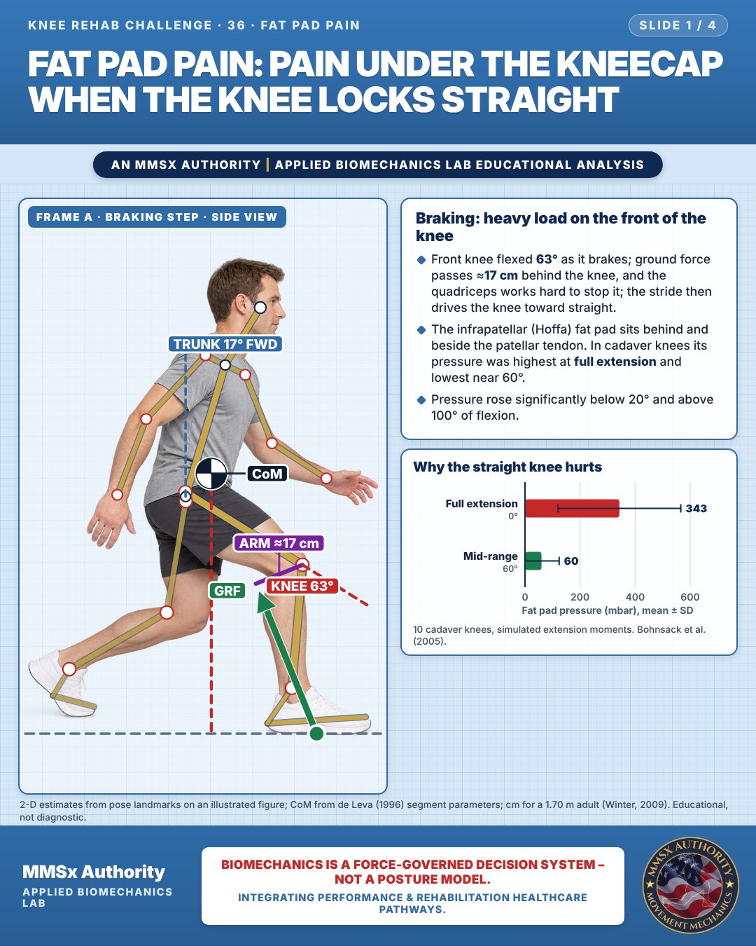
Braking: heavy load on the front of the knee
Front knee flexed 63° as it brakes; ground force passes ≈17 cm behind the knee, and the quadriceps works hard to stop it; the stride then drives the knee toward straight.
The infrapatellar (Hoffa) fat pad sits behind and beside the patellar tendon. In cadaver knees its pressure was highest at full extension and lowest near 60°.
Pressure rose significantly below 20° and above 100° of flexion.
Why the straight knee hurts
Movement B: Relaxed standing, true side view
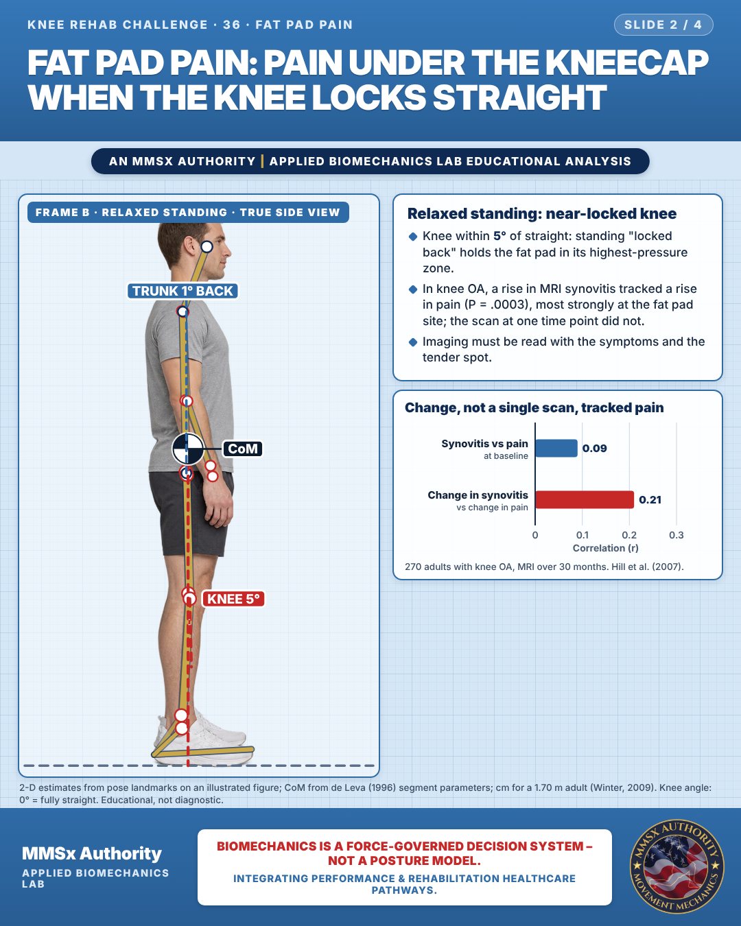
Relaxed standing: near-locked knee
Knee within 5° of straight: standing "locked back" holds the fat pad in its highest-pressure zone.
In knee OA, a rise in MRI synovitis tracked a rise in pain (P = .0003), most strongly at the fat pad site; the scan at one time point did not.
Imaging must be read with the symptoms and the tender spot.
Change, not a single scan, tracked pain
Movement C: Seated reference (front view)
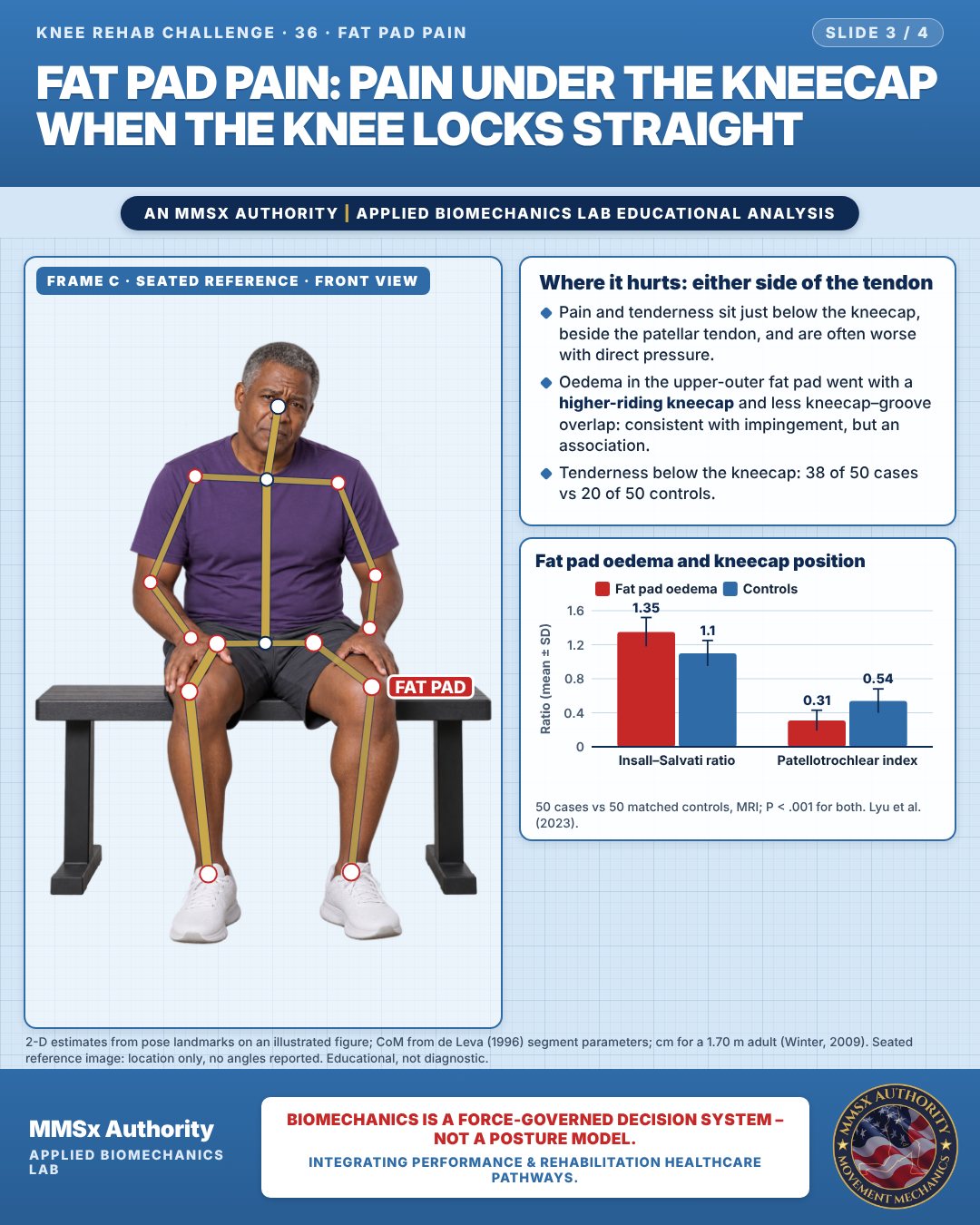
Where it hurts: either side of the tendon
Pain and tenderness sit just below the kneecap, beside the patellar tendon, and are often worse with direct pressure.
Oedema in the upper-outer fat pad went with a higher-riding kneecap and less kneecap–groove overlap: consistent with impingement, but an association.
Tenderness below the kneecap: 38 of 50 cases vs 20 of 50 controls.
Fat pad oedema and kneecap position
Decision pathway
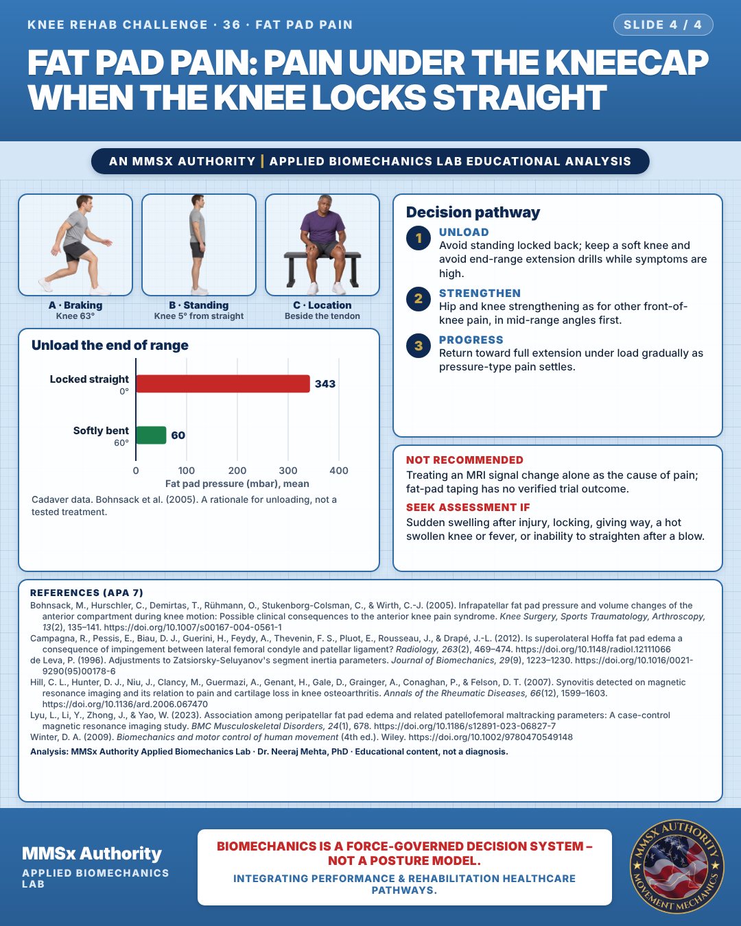
Unload the end of range
Unload
Avoid standing locked back; keep a soft knee and avoid end-range extension drills while symptoms are high.
Strengthen
Hip and knee strengthening as for other front-of-knee pain, in mid-range angles first.
Progress
Return toward full extension under load gradually as pressure-type pain settles.
What the evidence supports
- Not recommended: treating an MRI signal change alone as the source of pain — baseline synovitis did not correlate with baseline pain (Hill et al., 2007).
Myth and evidence
Myth“MRI shows fat pad inflammation, so that must be the cause of my pain.”
EvidenceAt baseline, synovitis (including the fat pad site) was not correlated with pain; it was change over time that tracked change in pain (Hill et al., 2007). Imaging must be interpreted alongside symptoms and the tender site.
Cautions
Not recommended
Treating an MRI signal change alone as the cause of pain; fat-pad taping has no verified trial outcome.
Seek assessment if
Sudden swelling after injury, locking, giving way, a hot swollen knee or fever, or inability to straighten after a blow.
Methods note
2-D estimates from pose landmarks on an illustrated figure; CoM from de Leva (1996) segment parameters; cm for a 1.70 m adult (Winter, 2009). Knee angle: 0° = fully straight. Seated reference image: location only, no angles reported. These are educational estimates, not a diagnosis.
References
- Bohnsack, M., Hurschler, C., Demirtas, T., Rühmann, O., Stukenborg-Colsman, C., & Wirth, C.-J. (2005). Infrapatellar fat pad pressure and volume changes of the anterior compartment during knee motion: Possible clinical consequences to the anterior knee pain syndrome. Knee Surgery, Sports Traumatology, Arthroscopy, 13(2), 135–141. https://doi.org/10.1007/s00167-004-0561-1
- Campagna, R., Pessis, E., Biau, D. J., Guerini, H., Feydy, A., Thevenin, F. S., Pluot, E., Rousseau, J., & Drapé, J.-L. (2012). Is superolateral Hoffa fat pad edema a consequence of impingement between lateral femoral condyle and patellar ligament? Radiology, 263(2), 469–474. https://doi.org/10.1148/radiol.12111066
- de Leva, P. (1996). Adjustments to Zatsiorsky-Seluyanov's segment inertia parameters. Journal of Biomechanics, 29(9), 1223–1230. https://doi.org/10.1016/0021-9290(95)00178-6
- Hill, C. L., Hunter, D. J., Niu, J., Clancy, M., Guermazi, A., Genant, H., Gale, D., Grainger, A., Conaghan, P., & Felson, D. T. (2007). Synovitis detected on magnetic resonance imaging and its relation to pain and cartilage loss in knee osteoarthritis. Annals of the Rheumatic Diseases, 66(12), 1599–1603. https://doi.org/10.1136/ard.2006.067470
- Lyu, L., Li, Y., Zhong, J., & Yao, W. (2023). Association among peripatellar fat pad edema and related patellofemoral maltracking parameters: A case-control magnetic resonance imaging study. BMC Musculoskeletal Disorders, 24(1), 678. https://doi.org/10.1186/s12891-023-06827-7
- Winter, D. A. (2009). Biomechanics and motor control of human movement (4th ed.). Wiley. https://doi.org/10.1002/9780470549148
Cite this article
Mehta, N. (2026, October 23). Fat pad pain: pain under the kneecap when the knee locks straight. MMSx Authority Applied Biomechanics Lab. https://mmsxauthority.org/edu/analysis/knee/36-fat-pad-pain-pain-under-the-kneecap-when-the-knee-locks-straight/
Part of the collection
Mehta, N. (2026). Knee Rehabilitation: 50 Evidence-Based Biomechanics Analyses [Collection]. MMSx Authority Applied Biomechanics Lab. Zenodo. https://doi.org/10.5281/zenodo.23735752