Knee Rehab Challenge · 35
Pes anserine pain: tenderness just below the inner knee
Pes anserine (anserine) pain syndrome is pain and local tenderness over the inner (medial) upper shin, about 3–5 cm below the medial joint line,…
Key points
- Confirm the site. Tenderness 3–5 cm below the inner joint line; check for knee OA, which usually travels with it.
- Treat within oa care. Exercise and weight management; physiotherapy and local injection both improved pain and function at 8 weeks, with no difference between them.
- Progress. Graded step-ups and stairs as the tender area settles.
What it is
Pes anserine (anserine) pain syndrome is pain and local tenderness over the inner (medial) upper shin, about 3–5 cm below the medial joint line, where the sartorius, gracilis and semitendinosus tendons insert and the anserine bursa lies beneath them; it is usually a clinical diagnosis, and imaging often does not show a distended bursa or tendon change.
How we analysed the movement
Three movements relevant to this topic were analysed from illustrated reference figures. Joint centres were located with two-dimensional pose estimation; joint angles, the whole-body centre of mass and, where relevant, the ground-reaction-force line and its moment arm about the knee were then calculated from standard segment parameters. The figures are educational estimates that show how position changes mechanical demand; they are not measurements of a patient. Every research value quoted below comes from the published source cited beside it.
Movement A: Stair ascent with rail (side view)
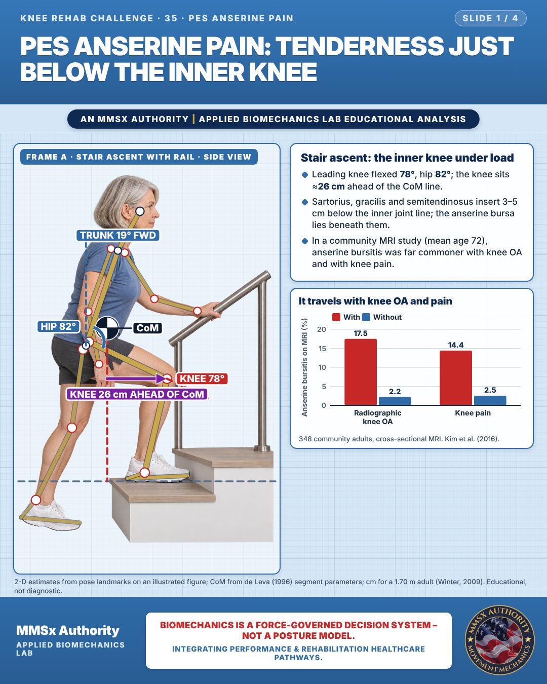
Stair ascent: the inner knee under load
Leading knee flexed 78°, hip 82°; the knee sits ≈26 cm ahead of the CoM line.
Sartorius, gracilis and semitendinosus insert 3–5 cm below the inner joint line; the anserine bursa lies beneath them.
In a community MRI study (mean age 72), anserine bursitis was far commoner with knee OA and with knee pain.
It travels with knee OA and pain
Movement B: Shallow squat (front view)
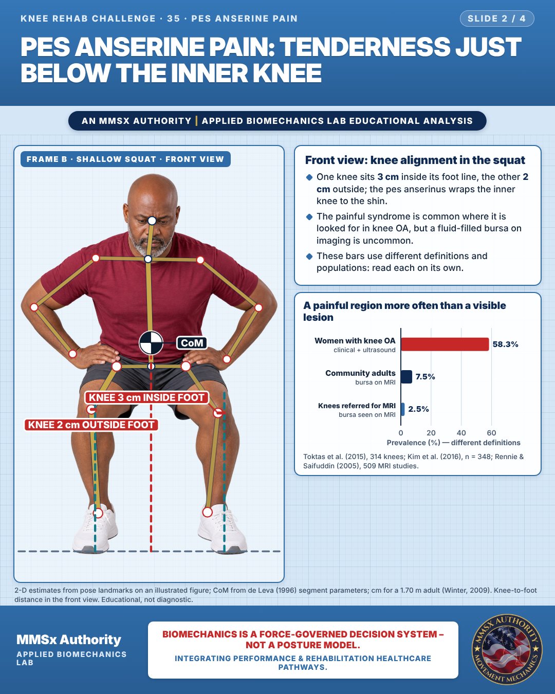
Front view: knee alignment in the squat
One knee sits 3 cm inside its foot line, the other 2 cm outside; the pes anserinus wraps the inner knee to the shin.
The painful syndrome is common where it is looked for in knee OA, but a fluid-filled bursa on imaging is uncommon.
These bars use different definitions and populations: read each on its own.
A painful region more often than a visible lesion
Movement C: Seated reference (front view)
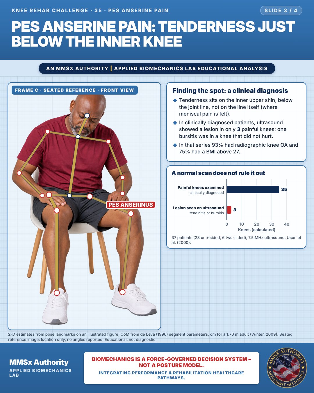
Finding the spot: a clinical diagnosis
Tenderness sits on the inner upper shin, below the joint line, not on the line itself (where meniscal pain is felt).
In clinically diagnosed patients, ultrasound showed a lesion in only 3 painful knees; one bursitis was in a knee that did not hurt.
In that series 93% had radiographic knee OA and 75% had a BMI above 27.
A normal scan does not rule it out
Decision pathway
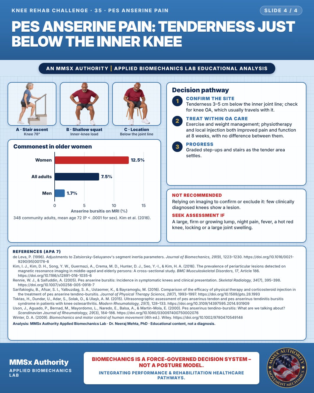
Commonest in older women
Confirm the site
Tenderness 3–5 cm below the inner joint line; check for knee OA, which usually travels with it.
Treat within oa care
Exercise and weight management; physiotherapy and local injection both improved pain and function at 8 weeks, with no difference between them.
Progress
Graded step-ups and stairs as the tender area settles.
What the evidence supports
- Because the syndrome usually co-exists with knee OA (Kim et al., 2016; Toktas et al., 2015), management should sit inside OA care: exercise and weight management where relevant, with local measures for the tender area.
Myth and evidence
Myth“It is a bursitis, so the scan will show it.”
EvidenceIn clinically diagnosed patients, ultrasound showed bursitis or tendinitis in only a few knees (Uson et al., 2000), and MRI identified anserine bursitis in only 2.5% of symptomatic knees referred for imaging (Rennie & Saifuddin, 2005). The diagnosis is clinical, by site of tenderness.
Cautions
Not recommended
Relying on imaging to confirm or exclude it: few clinically diagnosed knees show a lesion.
Seek assessment if
A large, firm or growing lump, night pain, fever, a hot red knee, locking or a large joint swelling.
Methods note
2-D estimates from pose landmarks on an illustrated figure; CoM from de Leva (1996) segment parameters; cm for a 1.70 m adult (Winter, 2009). Knee-to-foot distance in the front view. Seated reference image: location only, no angles reported. These are educational estimates, not a diagnosis.
References
- de Leva, P. (1996). Adjustments to Zatsiorsky-Seluyanov's segment inertia parameters. Journal of Biomechanics, 29(9), 1223–1230. https://doi.org/10.1016/0021-9290(95)00178-6
- Kim, I. J., Kim, D. H., Song, Y. W., Guermazi, A., Crema, M. D., Hunter, D. J., Seo, Y.-I., & Kim, H. A. (2016). The prevalence of periarticular lesions detected on magnetic resonance imaging in middle-aged and elderly persons: A cross-sectional study. BMC Musculoskeletal Disorders, 17, Article 186. https://doi.org/10.1186/s12891-016-1035-6
- Rennie, W. J., & Saifuddin, A. (2005). Pes anserine bursitis: Incidence in symptomatic knees and clinical presentation. Skeletal Radiology, 34(7), 395–398. https://doi.org/10.1007/s00256-005-0918-7
- Sarifakioglu, B., Afsar, S. I., Yalbuzdag, S. A., Ustaomer, K., & Bayramoglu, M. (2016). Comparison of the efficacy of physical therapy and corticosteroid injection in the treatment of pes anserine tendino-bursitis. Journal of Physical Therapy Science, 28(7), 1993–1997. https://doi.org/10.1589/jpts.28.1993
- Toktas, H., Dundar, U., Adar, S., Solak, O., & Ulaşlı, A. M. (2015). Ultrasonographic assessment of pes anserinus tendon and pes anserinus tendinitis bursitis syndrome in patients with knee osteoarthritis. Modern Rheumatology, 25(1), 128–133. https://doi.org/10.3109/14397595.2014.931909
- Uson, J., Aguado, P., Bernad, M., Mayordomo, L., Naredo, E., Balsa, A., & Martín-Mola, E. (2000). Pes anserinus tendino-bursitis: What are we talking about? Scandinavian Journal of Rheumatology, 29(3), 184–186. https://doi.org/10.1080/030097400750002076
- Winter, D. A. (2009). Biomechanics and motor control of human movement (4th ed.). Wiley. https://doi.org/10.1002/9780470549148
Cite this article
Mehta, N. (2026, October 23). Pes anserine pain: tenderness just below the inner knee. MMSx Authority Applied Biomechanics Lab. https://mmsxauthority.org/edu/analysis/knee/35-pes-anserine-pain-tenderness-just-below-the-inner-knee/
Part of the collection
Mehta, N. (2026). Knee Rehabilitation: 50 Evidence-Based Biomechanics Analyses [Collection]. MMSx Authority Applied Biomechanics Lab. Zenodo. https://doi.org/10.5281/zenodo.23735752