Knee Rehab Challenge · 33
Weak quadriceps: why thigh strength protects your knee
Quadriceps weakness is reduced knee-extensor force capacity relative to the other limb or to age- and sex-matched norms.
Key points
- Switch it on. Settle swelling and pain first; quadriceps activation work where inhibition is present.
- Strengthen. Progressive quadriceps strengthening: step-ups, split squats, chair rises, adding load over weeks.
- Keep going. Benefits shrank by 2–6 months after programmes ended: strength work needs to continue.
What it is
Quadriceps weakness is reduced knee-extensor force capacity relative to the other limb or to age- and sex-matched norms. It can come from muscle atrophy and/or failure of voluntary activation; when a swollen, painful or injured joint reflexly reduces quadriceps drive, this is called arthrogenic muscle inhibition (AMI).
How we analysed the movement
Three movements relevant to this topic were analysed from illustrated reference figures. Joint centres were located with two-dimensional pose estimation; joint angles, the whole-body centre of mass and, where relevant, the ground-reaction-force line and its moment arm about the knee were then calculated from standard segment parameters. The figures are educational estimates that show how position changes mechanical demand; they are not measurements of a patient. Every research value quoted below comes from the published source cited beside it.
Movement A: Step-up (side view)
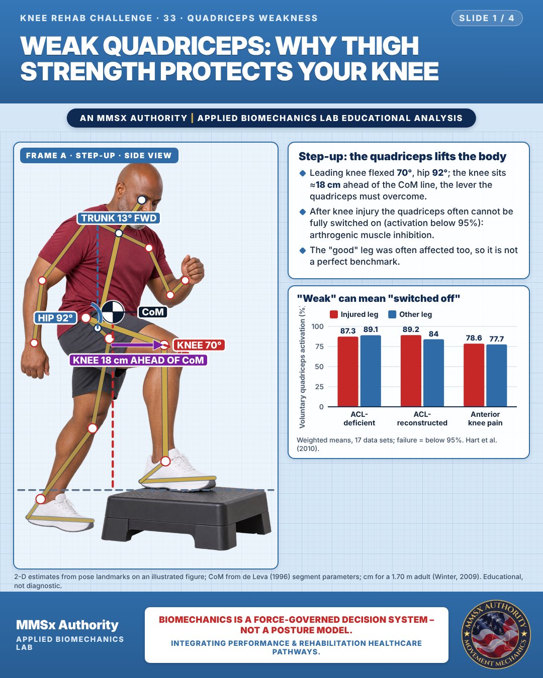
Step-up: the quadriceps lifts the body
Leading knee flexed 70°, hip 92°; the knee sits ≈18 cm ahead of the CoM line, the lever the quadriceps must overcome.
After knee injury the quadriceps often cannot be fully switched on (activation below 95%): arthrogenic muscle inhibition.
The "good" leg was often affected too, so it is not a perfect benchmark.
"Weak" can mean "switched off"
Movement B: Split squat (front view)
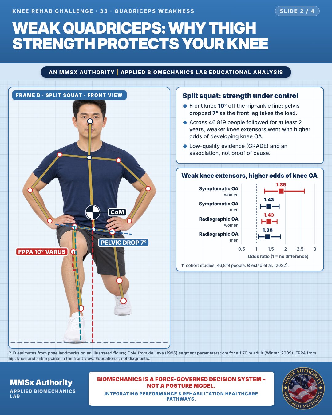
Split squat: strength under control
Front knee 10° off the hip–ankle line; pelvis dropped 7° as the front leg takes the load.
Across 46,819 people followed for at least 2 years, weaker knee extensors went with higher odds of developing knee OA.
Low-quality evidence (GRADE) and an association, not proof of cause.
Weak knee extensors, higher odds of knee OA
Movement C: Rising from a chair (side view)
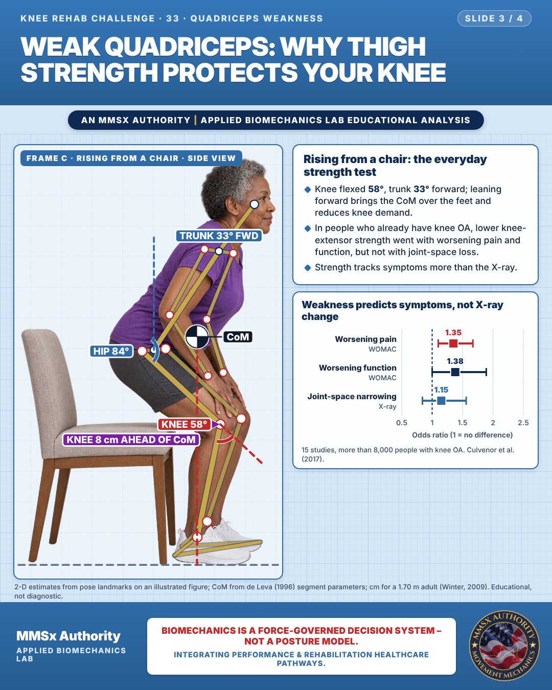
Rising from a chair: the everyday strength test
Knee flexed 58°, trunk 33° forward; leaning forward brings the CoM over the feet and reduces knee demand.
In people who already have knee OA, lower knee-extensor strength went with worsening pain and function, but not with joint-space loss.
Strength tracks symptoms more than the X-ray.
Weakness predicts symptoms, not X-ray change
Decision pathway
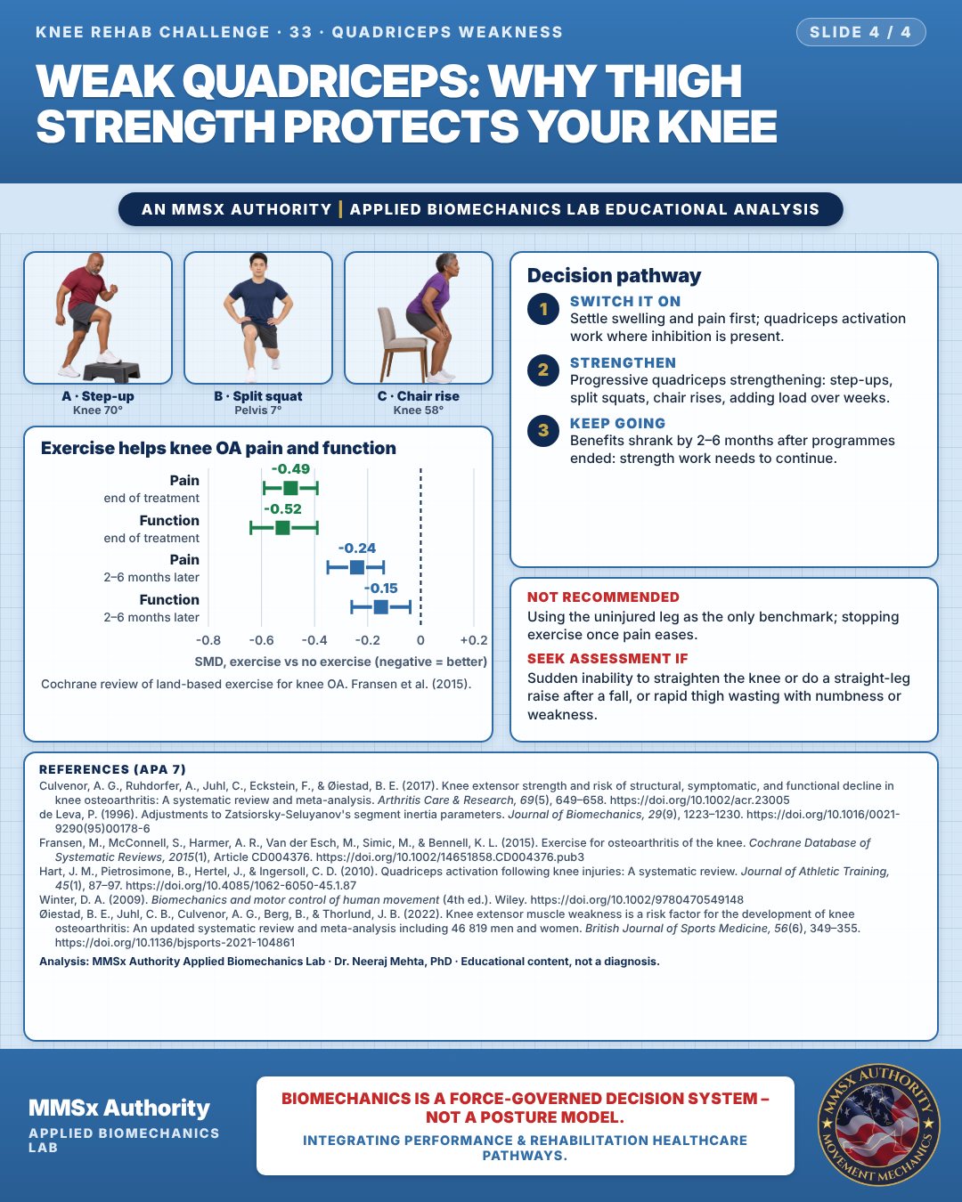
Exercise helps knee OA pain and function
Switch it on
Settle swelling and pain first; quadriceps activation work where inhibition is present.
Strengthen
Progressive quadriceps strengthening: step-ups, split squats, chair rises, adding load over weeks.
Keep going
Benefits shrank by 2–6 months after programmes ended: strength work needs to continue.
What the evidence supports
- Exercise therapy is recommended for knee OA and produces moderate short-term improvements in pain and function (Fransen et al., 2015).
- Quadriceps strengthening is a logical target because weakness is associated with incident OA (Øiestad et al., 2022) and with symptomatic/functional decline (Culvenor et al., 2017) — association, not proven cause.
- Not recommended: relying on the "good" leg as the only benchmark (bilateral deficits are common — Hart et al., 2010).
Myth and evidence
Myth“Strength work wears out the knee.”
EvidenceExercise improved pain and function vs no exercise, with withdrawals no higher than controls (Fransen et al., 2015).
Cautions
Not recommended
Using the uninjured leg as the only benchmark; stopping exercise once pain eases.
Seek assessment if
Sudden inability to straighten the knee or do a straight-leg raise after a fall, or rapid thigh wasting with numbness or weakness.
Methods note
2-D estimates from pose landmarks on an illustrated figure; CoM from de Leva (1996) segment parameters; cm for a 1.70 m adult (Winter, 2009). FPPA from hip, knee and ankle points in the front view. These are educational estimates, not a diagnosis.
References
- Culvenor, A. G., Ruhdorfer, A., Juhl, C., Eckstein, F., & Øiestad, B. E. (2017). Knee extensor strength and risk of structural, symptomatic, and functional decline in knee osteoarthritis: A systematic review and meta-analysis. Arthritis Care & Research, 69(5), 649–658. https://doi.org/10.1002/acr.23005
- de Leva, P. (1996). Adjustments to Zatsiorsky-Seluyanov's segment inertia parameters. Journal of Biomechanics, 29(9), 1223–1230. https://doi.org/10.1016/0021-9290(95)00178-6
- Fransen, M., McConnell, S., Harmer, A. R., Van der Esch, M., Simic, M., & Bennell, K. L. (2015). Exercise for osteoarthritis of the knee. Cochrane Database of Systematic Reviews, 2015(1), Article CD004376. https://doi.org/10.1002/14651858.CD004376.pub3
- Hart, J. M., Pietrosimone, B., Hertel, J., & Ingersoll, C. D. (2010). Quadriceps activation following knee injuries: A systematic review. Journal of Athletic Training, 45(1), 87–97. https://doi.org/10.4085/1062-6050-45.1.87
- Winter, D. A. (2009). Biomechanics and motor control of human movement (4th ed.). Wiley. https://doi.org/10.1002/9780470549148
- Øiestad, B. E., Juhl, C. B., Culvenor, A. G., Berg, B., & Thorlund, J. B. (2022). Knee extensor muscle weakness is a risk factor for the development of knee osteoarthritis: An updated systematic review and meta-analysis including 46 819 men and women. British Journal of Sports Medicine, 56(6), 349–355. https://doi.org/10.1136/bjsports-2021-104861
Cite this article
Mehta, N. (2026, October 23). Weak quadriceps: why thigh strength protects your knee. MMSx Authority Applied Biomechanics Lab. https://mmsxauthority.org/edu/analysis/knee/33-weak-quadriceps-why-thigh-strength-protects-your-knee/
Part of the collection
Mehta, N. (2026). Knee Rehabilitation: 50 Evidence-Based Biomechanics Analyses [Collection]. MMSx Authority Applied Biomechanics Lab. Zenodo. https://doi.org/10.5281/zenodo.23735752