Knee Rehab Challenge · 27
Hip and trunk control: how the whole body influences knee loading
"Hip and trunk control" describes how well the pelvis and trunk are held over the stance limb in the frontal and transverse planes.
Key points
- Assess the chain. Watch pelvis and trunk, not just the knee, in single-leg tasks and cuts.
- Train hip + knee. Combined hip and knee exercise is preferred over knee exercise alone (Collins et al., 2018).
- Coach technique. Narrower plant, trunk over the stance leg, less knee valgus when cutting; trunk-lean gait for OA remains experimental.
What it is
"Hip and trunk control" describes how well the pelvis and trunk are held over the stance limb in the frontal and transverse planes; where the trunk and pelvis sit changes the line of the ground reaction force relative to the knee and therefore the knee's frontal-plane (abduction/adduction) moment.
How we analysed the movement
Three movements relevant to this topic were analysed from illustrated reference figures. Joint centres were located with two-dimensional pose estimation; joint angles, the whole-body centre of mass and, where relevant, the ground-reaction-force line and its moment arm about the knee were then calculated from standard segment parameters. The figures are educational estimates that show how position changes mechanical demand; they are not measurements of a patient. Every research value quoted below comes from the published source cited beside it.
Movement A: Single-leg stance (rear view)
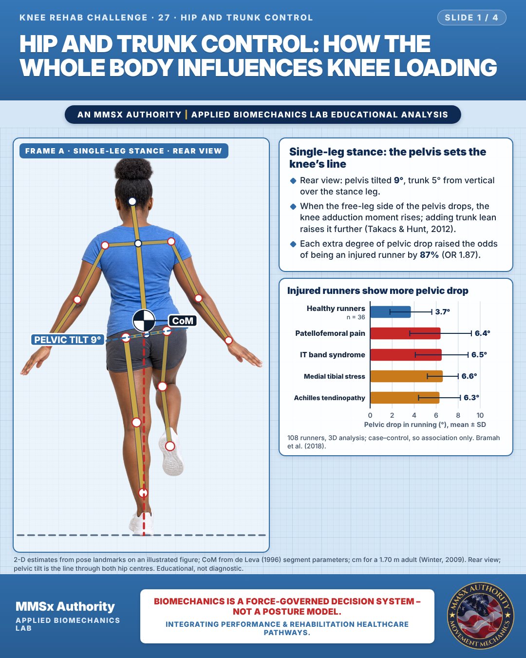
Single-leg stance: the pelvis sets the knee’s line
Rear view: pelvis tilted 9°, trunk 5° from vertical over the stance leg.
When the free-leg side of the pelvis drops, the knee adduction moment rises; adding trunk lean raises it further (Takacs & Hunt, 2012).
Each extra degree of pelvic drop raised the odds of being an injured runner by 87% (OR 1.87).
Injured runners show more pelvic drop
Movement B: Single-leg squat (front view)
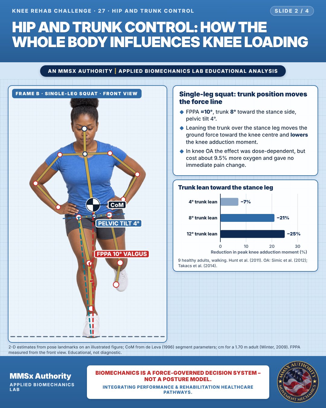
Single-leg squat: trunk position moves the force line
FPPA ≈10°, trunk 8° toward the stance side, pelvic tilt 4°.
Leaning the trunk over the stance leg moves the ground force toward the knee centre and lowers the knee adduction moment.
In knee OA the effect was dose-dependent, but cost about 9.5% more oxygen and gave no immediate pain change.
Trunk lean toward the stance leg
Movement C: Direction change, oblique
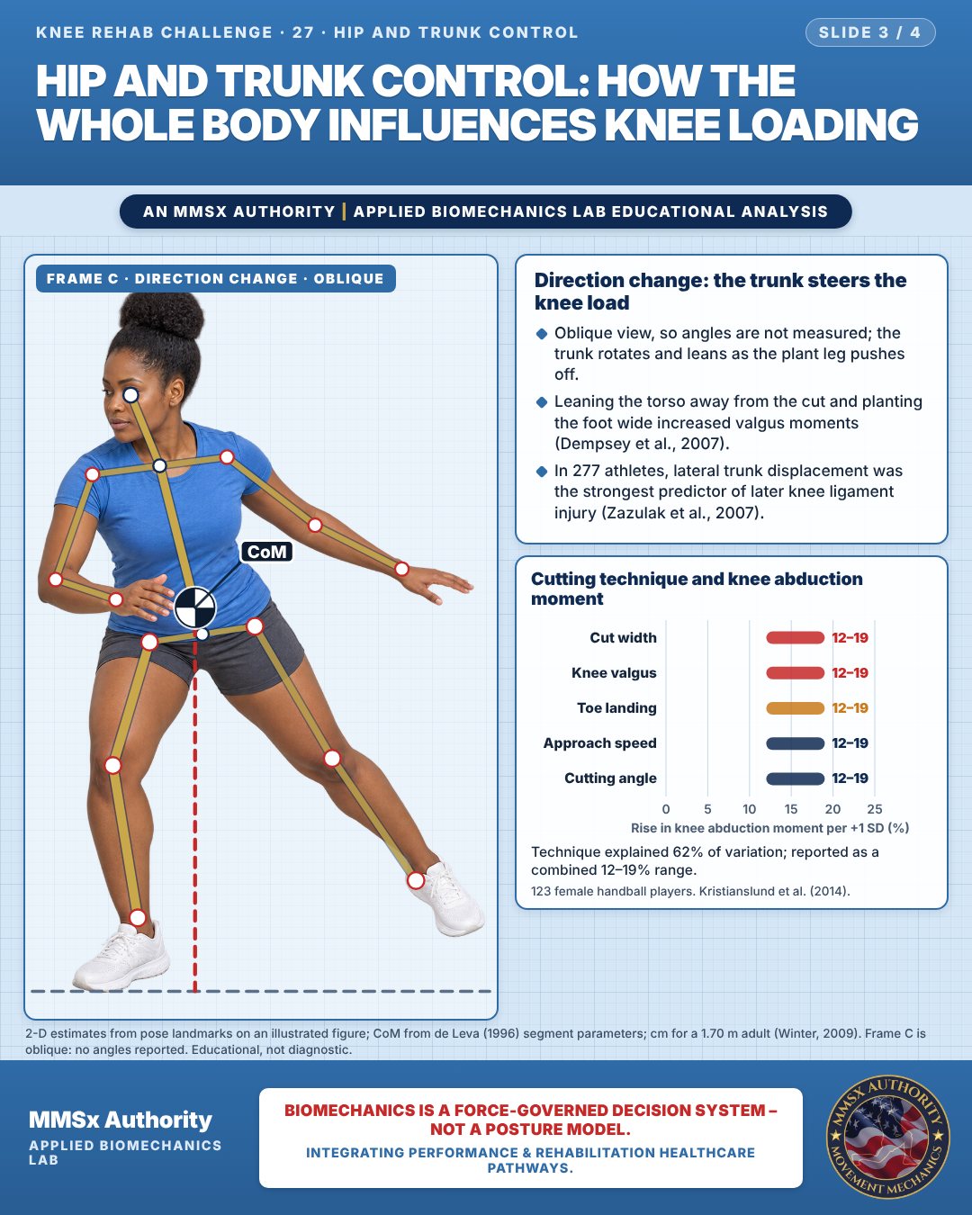
Direction change: the trunk steers the knee load
Oblique view, so angles are not measured; the trunk rotates and leans as the plant leg pushes off.
Leaning the torso away from the cut and planting the foot wide increased valgus moments (Dempsey et al., 2007).
In 277 athletes, lateral trunk displacement was the strongest predictor of later knee ligament injury (Zazulak et al., 2007).
Cutting technique and knee abduction moment
Technique explained 62% of variation; reported as a combined 12–19% range.
Decision pathway
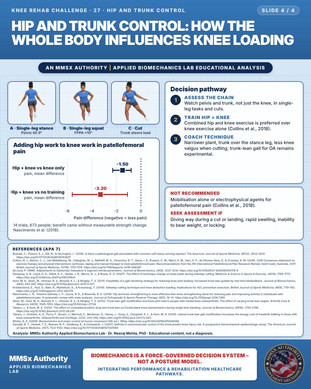
Adding hip work to knee work in patellofemoral pain
Assess the chain
Watch pelvis and trunk, not just the knee, in single-leg tasks and cuts.
Train hip + knee
Combined hip and knee exercise is preferred over knee exercise alone (Collins et al., 2018).
Coach technique
Narrower plant, trunk over the stance leg, less knee valgus when cutting; trunk-lean gait for OA remains experimental.
What the evidence supports
- Recommended (PFP): exercise therapy; combined hip and knee exercise preferred over knee exercise alone; exercise combined with foot orthoses/taping/manual therapy for short–medium-term pain; prefabricated foot orthoses short-term. Not recommended: mobilisation alone; electrophysical agents. Uncertain: gait retraining, taping/bracing, dry needling. (Collins et al., 2018)
- Knee OA: trunk-lean gait modification lowers the knee adduction moment in the laboratory but showed no immediate symptom change and raises the energy cost of walking (≈+9.5% VO2) — present as an experimental strategy, not a routine recommendation. (Simic et al., 2012; Takacs et al., 2014)
- ACL injury-risk reduction: technique factors (narrower cut, less knee valgus, trunk control) are modifiable targets identified by cross-sectional and prospective studies (Kristianslund et al., 2014; Zazulak et al., 2007).
Myth and evidence
Myth“Knee pain means you should only train the knee.”
EvidenceHip + knee strengthening reduced pain more than knee strengthening alone (MD −1.5, 95% CI −2.3 to −0.8) (Nascimento et al., 2018), and consensus prefers combined hip and knee exercise (Collins et al., 2018).
Myth“Hip exercise works by making the hip stronger.”
EvidenceBenefits occurred without significant measured strength change (Nascimento et al., 2018) — mechanisms likely include control/load tolerance, not strength alone.
Cautions
Not recommended
Mobilisation alone or electrophysical agents for patellofemoral pain (Collins et al., 2018).
Seek assessment if
Giving way during a cut or landing, rapid swelling, inability to bear weight, or locking.
Methods note
2-D estimates from pose landmarks on an illustrated figure; CoM from de Leva (1996) segment parameters; cm for a 1.70 m adult (Winter, 2009). Rear view; pelvic tilt is the line through both hip centres. FPPA measured from the front view. Frame C is oblique: no angles reported. These are educational estimates, not a diagnosis.
References
- Bramah, C., Preece, S. J., Gill, N., & Herrington, L. (2018). Is there a pathological gait associated with common soft tissue running injuries? The American Journal of Sports Medicine, 46(12), 3023–3031. https://doi.org/10.1177/0363546518793657
- Collins, N. J., Barton, C. J., van Middelkoop, M., Callaghan, M. J., Rathleff, M. S., Vicenzino, B. T., Davis, I. S., Powers, C. M., Macri, E. M., Hart, H. F., de Oliveira Silva, D., & Crossley, K. M. (2018). 2018 Consensus statement on exercise therapy and physical interventions (orthoses, taping and manual therapy) to treat patellofemoral pain: Recommendations from the 5th International Patellofemoral Pain Research Retreat, Gold Coast, Australia, 2017. British Journal of Sports Medicine, 52(18), 1170–1178. https://doi.org/10.1136/bjsports-2018-099397
- de Leva, P. (1996). Adjustments to Zatsiorsky-Seluyanov's segment inertia parameters. Journal of Biomechanics, 29(9), 1223–1230. https://doi.org/10.1016/0021-9290(95)00178-6
- Dempsey, A. R., Lloyd, D. G., Elliott, B. C., Steele, J. R., Munro, B. J., & Russo, K. A. (2007). The effect of technique change on knee loads during sidestep cutting. Medicine & Science in Sports & Exercise, 39(10), 1765–1773. https://doi.org/10.1249/mss.0b013e31812f56d1
- Hunt, M. A., Simic, M., Hinman, R. S., Bennell, K. L., & Wrigley, T. V. (2011). Feasibility of a gait retraining strategy for reducing knee joint loading: Increased trunk lean guided by real-time biofeedback. Journal of Biomechanics, 44(5), 943–947. https://doi.org/10.1016/j.jbiomech.2010.11.027
- Kristianslund, E., Faul, O., Bahr, R., Myklebust, G., & Krosshaug, T. (2014). Sidestep cutting technique and knee abduction loading: Implications for ACL prevention exercises. British Journal of Sports Medicine, 48(9), 779–783. https://doi.org/10.1136/bjsports-2012-091370
- Nascimento, L. R., Teixeira-Salmela, L. F., Souza, R. B., & Resende, R. A. (2018). Hip and knee strengthening is more effective than knee strengthening alone for reducing pain and improving activity in individuals with patellofemoral pain: A systematic review with meta-analysis. Journal of Orthopaedic & Sports Physical Therapy, 48(1), 19–31. https://doi.org/10.2519/jospt.2018.7365
- Simic, M., Hunt, M. A., Bennell, K. L., Hinman, R. S., & Wrigley, T. V. (2012). Trunk lean gait modification and knee joint load in people with medial knee osteoarthritis: The effect of varying trunk lean angles. Arthritis Care & Research, 64(10), 1545–1553. https://doi.org/10.1002/acr.21724
- Takacs, J., & Hunt, M. A. (2012). The effect of contralateral pelvic drop and trunk lean on frontal plane knee biomechanics during single limb standing. Journal of Biomechanics, 45(16), 2791–2796. https://doi.org/10.1016/j.jbiomech.2012.08.041
- Takacs, J., Kirkham, A. A., Perry, F., Brown, J., Marriott, E., Monkman, D., Havey, J., Hung, S., Campbell, K. L., & Hunt, M. A. (2014). Lateral trunk lean gait modification increases the energy cost of treadmill walking in those with knee osteoarthritis. Osteoarthritis and Cartilage, 22(2), 203–209. https://doi.org/10.1016/j.joca.2013.12.003
- Winter, D. A. (2009). Biomechanics and motor control of human movement (4th ed.). Wiley. https://doi.org/10.1002/9780470549148
- Zazulak, B. T., Hewett, T. E., Reeves, N. P., Goldberg, B., & Cholewicki, J. (2007). Deficits in neuromuscular control of the trunk predict knee injury risk: A prospective biomechanical-epidemiologic study. The American Journal of Sports Medicine, 35(7), 1123–1130. https://doi.org/10.1177/0363546507301585
Cite this article
Mehta, N. (2026, October 23). Hip and trunk control: how the whole body influences knee loading. MMSx Authority Applied Biomechanics Lab. https://mmsxauthority.org/edu/analysis/knee/27-hip-and-trunk-control-how-the-whole-body-influences-knee-loading/
Part of the collection
Mehta, N. (2026). Knee Rehabilitation: 50 Evidence-Based Biomechanics Analyses [Collection]. MMSx Authority Applied Biomechanics Lab. Zenodo. https://doi.org/10.5281/zenodo.23735752