Knee Rehab Challenge · 25
Knees moving inward: what does dynamic knee valgus tell us?
Dynamic knee valgus is the visible inward movement of the knee relative to the hip and foot during a weight-bearing task.
Key points
- Observe. Film consistently (same camera height, distance, task); treat changes under ~8° with caution.
- Interpret. Valgus is one observation within the clinical picture, not a screening verdict on its own.
- Train. Neuromuscular prevention programmes for sport; hip plus knee exercise for patellofemoral pain (Crossley et al., 2016).
What it is
Dynamic knee valgus is the visible inward movement of the knee relative to the hip and foot during a weight-bearing task; it is a combination of hip adduction/internal rotation, knee abduction and tibial/foot motion, not a pure "knee" movement. In clinics it is usually measured on 2D video as the frontal-plane projection angle (FPPA). It is a movement observation associated (not deterministically) with patellofemoral pain and, in some studies, ACL injury risk.
How we analysed the movement
Three movements relevant to this topic were analysed from illustrated reference figures. Joint centres were located with two-dimensional pose estimation; joint angles, the whole-body centre of mass and, where relevant, the ground-reaction-force line and its moment arm about the knee were then calculated from standard segment parameters. The figures are educational estimates that show how position changes mechanical demand; they are not measurements of a patient. Every research value quoted below comes from the published source cited beside it.
Movement A: Single-leg squat (front view)
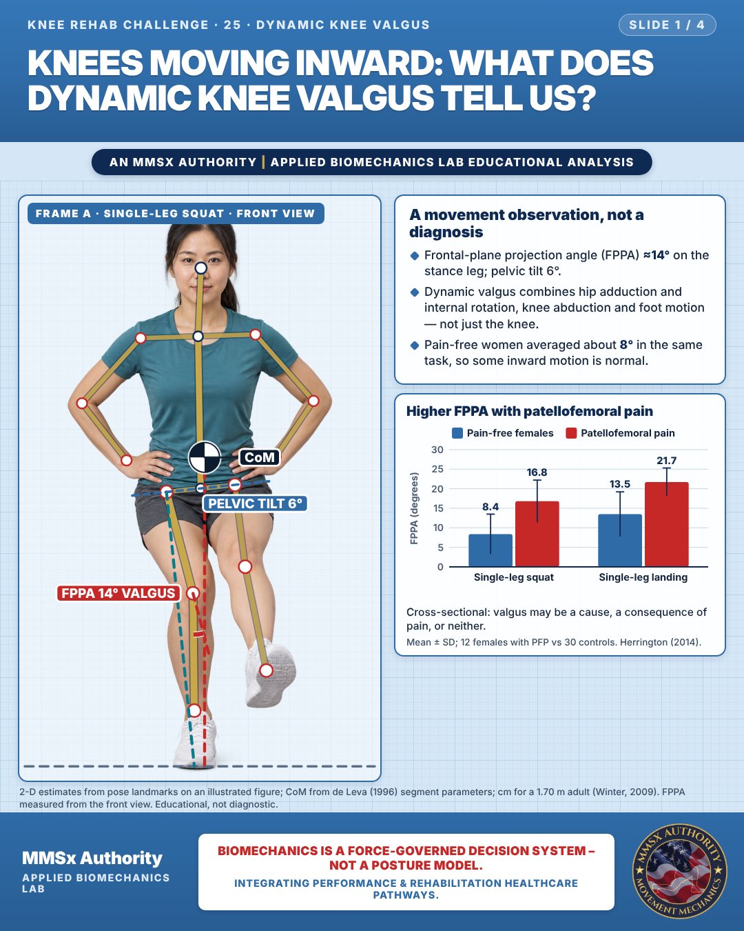
A movement observation, not a diagnosis
Frontal-plane projection angle (FPPA) ≈14° on the stance leg; pelvic tilt 6°.
Dynamic valgus combines hip adduction and internal rotation, knee abduction and foot motion — not just the knee.
Pain-free women averaged about 8° in the same task, so some inward motion is normal.
Higher FPPA with patellofemoral pain
Cross-sectional: valgus may be a cause, a consequence of pain, or neither.
Movement B: Step-down (front view)
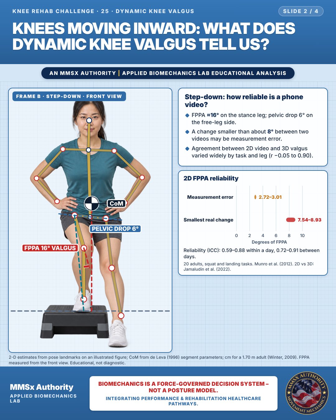
Step-down: how reliable is a phone video?
FPPA ≈16° on the stance leg; pelvic drop 6° on the free-leg side.
A change smaller than about 8° between two videos may be measurement error.
Agreement between 2D video and 3D valgus varied widely by task and leg (r −0.05 to 0.90).
2D FPPA reliability
Reliability (ICC): 0.59–0.88 within a day, 0.72–0.91 between days.
Movement C: Landing, oblique
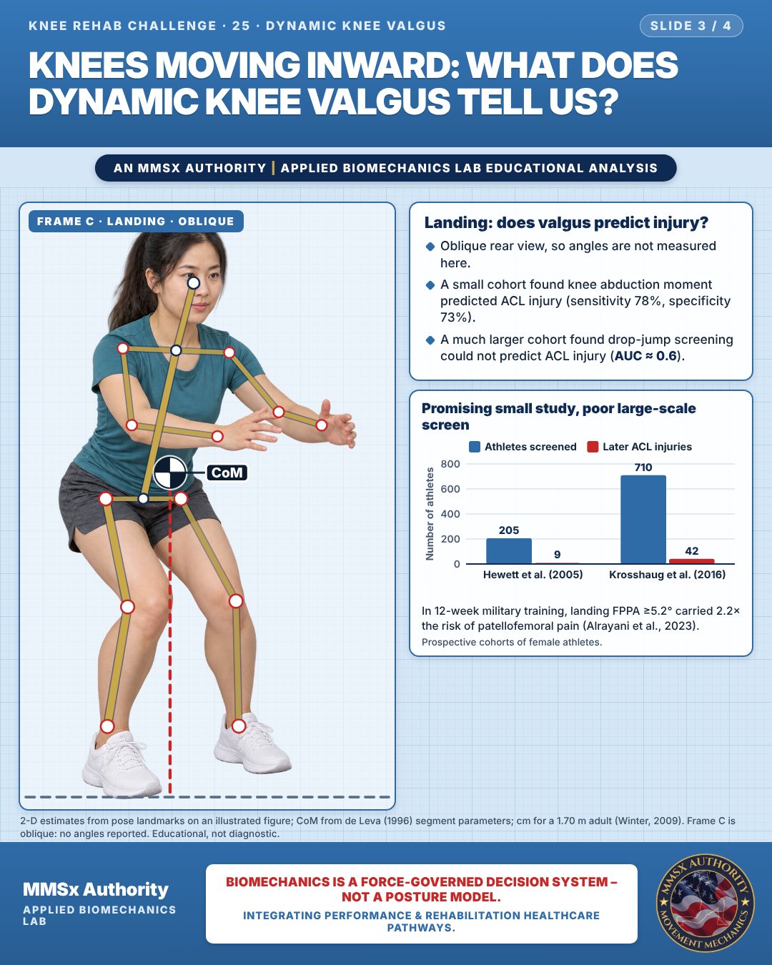
Landing: does valgus predict injury?
Oblique rear view, so angles are not measured here.
A small cohort found knee abduction moment predicted ACL injury (sensitivity 78%, specificity 73%).
A much larger cohort found drop-jump screening could not predict ACL injury (AUC ≈ 0.6).
Promising small study, poor large-scale screen
In 12-week military training, landing FPPA ≥5.2° carried 2.2× the risk of patellofemoral pain (Alrayani et al., 2023).
Decision pathway
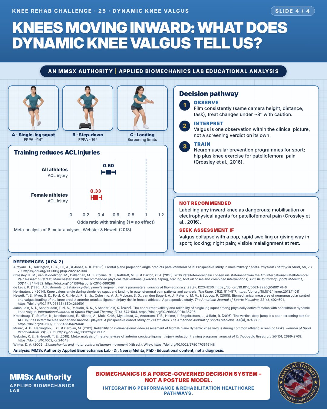
Training reduces ACL injuries
Observe
Film consistently (same camera height, distance, task); treat changes under ~8° with caution.
Interpret
Valgus is one observation within the clinical picture, not a screening verdict on its own.
Train
Neuromuscular prevention programmes for sport; hip plus knee exercise for patellofemoral pain (Crossley et al., 2016).
What the evidence supports
- Use dynamic knee valgus as one observation within a clinical picture — not as a stand-alone screening verdict (Krosshaug et al., 2016: AUC ≈ 0.6).
- For ACL risk reduction: structured neuromuscular/injury-prevention training programmes (≈50% overall, ≈67% in females; Webster & Hewett, 2018).
- Measurement decisions: film consistently; treat changes < ~8° FPPA cautiously (SDD 7.54–8.93°; Munro et al., 2012).
Myth and evidence
Myth“Any knee that moves inward is dangerous and will tear the ACL.”
EvidenceThe larger prospective cohort found drop-jump screening could not predict ACL injury (AUC ≈ 0.6; no association in previously uninjured players) (Krosshaug et al., 2016), and 2D FPPA has an SDD of ~8° (Munro et al., 2012). Healthy females showed a mean single-leg-squat FPPA of 8.4 ± 5.1° (Herrington, 2014) — some inward motion is normal. Valgus is an associated, modifiable observation, not a diagnosis.
Cautions
Not recommended
Labelling any inward knee as dangerous; mobilisation or electrophysical agents for patellofemoral pain (Crossley et al., 2016).
Seek assessment if
Valgus collapse with a pop, rapid swelling or giving way in sport; locking; night pain; visible malalignment at rest.
Methods note
2-D estimates from pose landmarks on an illustrated figure; CoM from de Leva (1996) segment parameters; cm for a 1.70 m adult (Winter, 2009). FPPA measured from the front view. Frame C is oblique: no angles reported. These are educational estimates, not a diagnosis.
References
- Alrayani, H., Herrington, L. C., Liu, A., & Jones, R. K. (2023). Frontal plane projection angle predicts patellofemoral pain: Prospective study in male military cadets. Physical Therapy in Sport, 59, 73–79. https://doi.org/10.1016/j.ptsp.2022.12.004
- Crossley, K. M., van Middelkoop, M., Callaghan, M. J., Collins, N. J., Rathleff, M. S., & Barton, C. J. (2016). 2016 Patellofemoral pain consensus statement from the 4th International Patellofemoral Pain Research Retreat, Manchester. Part 2: Recommended physical interventions (exercise, taping, bracing, foot orthoses and combined interventions). British Journal of Sports Medicine, 50(14), 844–852. https://doi.org/10.1136/bjsports-2016-096268
- de Leva, P. (1996). Adjustments to Zatsiorsky-Seluyanov's segment inertia parameters. Journal of Biomechanics, 29(9), 1223–1230. https://doi.org/10.1016/0021-9290(95)00178-6
- Herrington, L. (2014). Knee valgus angle during single leg squat and landing in patellofemoral pain patients and controls. The Knee, 21(2), 514–517. https://doi.org/10.1016/j.knee.2013.11.011
- Hewett, T. E., Myer, G. D., Ford, K. R., Heidt, R. S., Jr., Colosimo, A. J., McLean, S. G., van den Bogert, A. J., Paterno, M. V., & Succop, P. (2005). Biomechanical measures of neuromuscular control and valgus loading of the knee predict anterior cruciate ligament injury risk in female athletes: A prospective study. The American Journal of Sports Medicine, 33(4), 492–501. https://doi.org/10.1177/0363546504269591
- Jamaludin, N. I., Sahabuddin, F. N. A., Rasudin, N. S., & Shaharudin, S. (2022). The concurrent validity and reliability of single leg squat among physically active females with and without dynamic knee valgus. International Journal of Sports Physical Therapy, 17(4), 574–584. https://doi.org/10.26603/001c.35706
- Krosshaug, T., Steffen, K., Kristianslund, E., Nilstad, A., Mok, K.-M., Myklebust, G., Andersen, T. E., Holme, I., Engebretsen, L., & Bahr, R. (2016). The vertical drop jump is a poor screening test for ACL injuries in female elite soccer and handball players: A prospective cohort study of 710 athletes. The American Journal of Sports Medicine, 44(4), 874–883. https://doi.org/10.1177/0363546515625048
- Munro, A. G., Herrington, L. C., & Carolan, M. (2012). Reliability of 2-dimensional video assessment of frontal-plane dynamic knee valgus during common athletic screening tasks. Journal of Sport Rehabilitation, 21(1), 7–11. https://doi.org/10.1123/jsr.21.1.7
- Webster, K. E., & Hewett, T. E. (2018). Meta-analysis of meta-analyses of anterior cruciate ligament injury reduction training programs. Journal of Orthopaedic Research, 36(10), 2696–2708. https://doi.org/10.1002/jor.24043
- Winter, D. A. (2009). Biomechanics and motor control of human movement (4th ed.). Wiley. https://doi.org/10.1002/9780470549148
Cite this article
Mehta, N. (2026, October 23). Knees moving inward: what does dynamic knee valgus tell us? MMSx Authority Applied Biomechanics Lab. https://mmsxauthority.org/edu/analysis/knee/25-knees-moving-inward-what-does-dynamic-knee-valgus-tell-us/
Part of the collection
Mehta, N. (2026). Knee Rehabilitation: 50 Evidence-Based Biomechanics Analyses [Collection]. MMSx Authority Applied Biomechanics Lab. Zenodo. https://doi.org/10.5281/zenodo.23735752