Knee Rehab Challenge · 22
Knee pain during lunges: depth, position, and load
Lunge-provoked knee pain is usually anterior (patellofemoral or patellar-tendon region) pain that rises with knee-flexion depth, step geometry and…
Key points
- Depth. Start in a pain-acceptable range (often under 50–60°) and extend depth gradually.
- Geometry. Use a longer step and no forward stride; try reverse lunges first.
- Load. Master bodyweight before adding external load — loaded lunges multiply kneecap force.
What it is
Lunge-provoked knee pain is usually anterior (patellofemoral or patellar-tendon region) pain that rises with knee-flexion depth, step geometry and external load; patellofemoral joint force and stress increase as the knee bends further under load, so depth, step length and lunge direction are adjustable "dose" variables rather than fixed rights or wrongs.
How we analysed the movement
Three movements relevant to this topic were analysed from illustrated reference figures. Joint centres were located with two-dimensional pose estimation; joint angles, the whole-body centre of mass and, where relevant, the ground-reaction-force line and its moment arm about the knee were then calculated from standard segment parameters. The figures are educational estimates that show how position changes mechanical demand; they are not measurements of a patient. Every research value quoted below comes from the published source cited beside it.
Movement A: Forward lunge (side view)
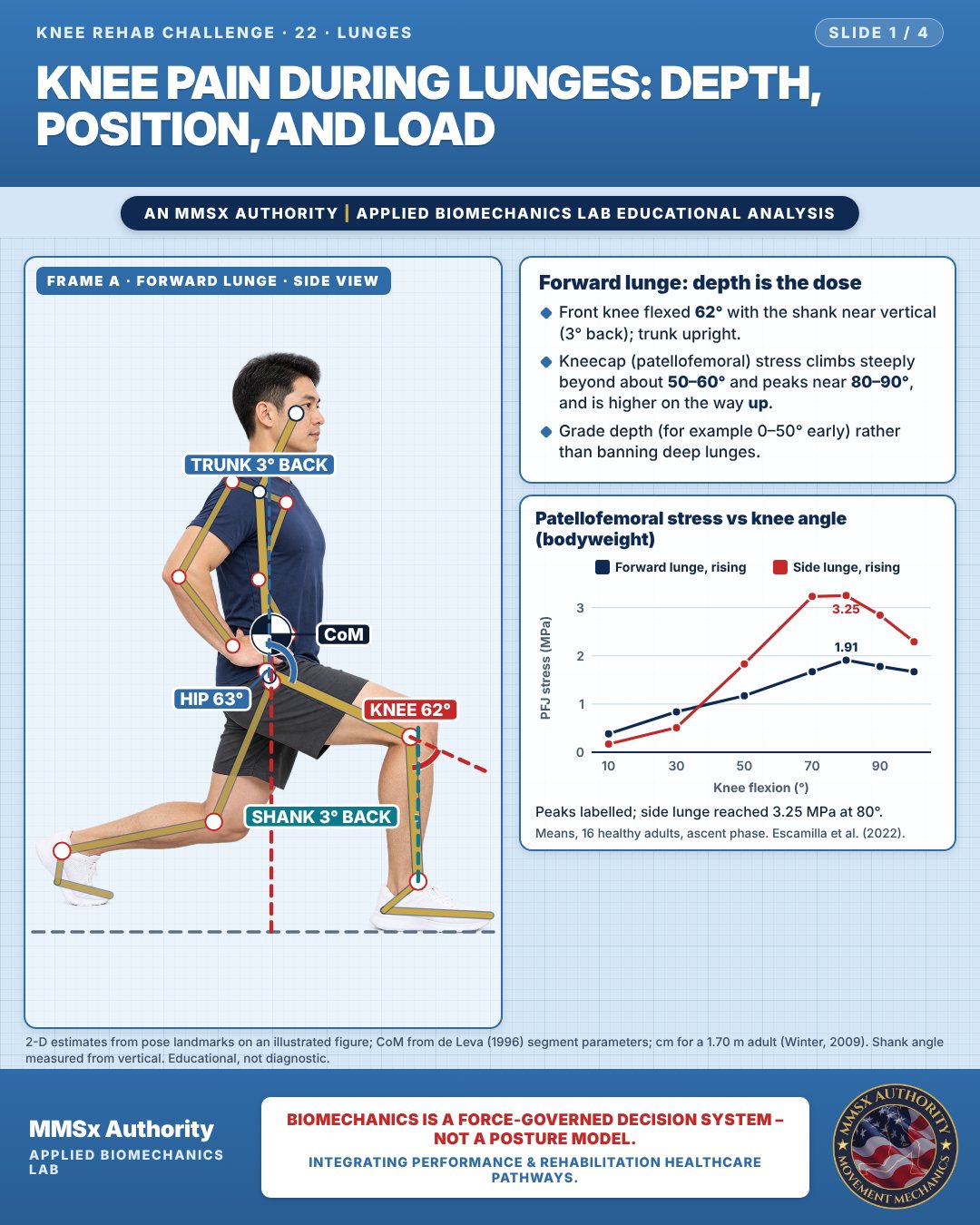
Forward lunge: depth is the dose
Front knee flexed 62° with the shank near vertical (3° back); trunk upright.
Kneecap (patellofemoral) stress climbs steeply beyond about 50–60° and peaks near 80–90°, and is higher on the way up.
Grade depth (for example 0–50° early) rather than banning deep lunges.
Patellofemoral stress vs knee angle (bodyweight)
Peaks labelled; side lunge reached 3.25 MPa at 80°.
Movement B: Split squat (front view)
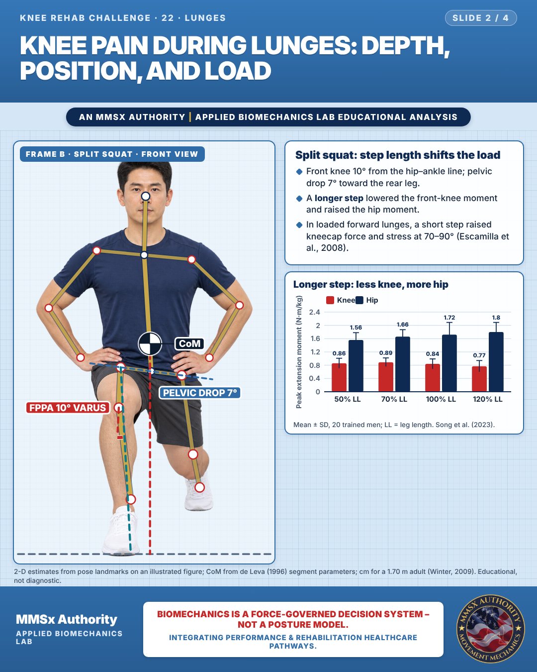
Split squat: step length shifts the load
Front knee 10° from the hip–ankle line; pelvic drop 7° toward the rear leg.
A longer step lowered the front-knee moment and raised the hip moment.
In loaded forward lunges, a short step raised kneecap force and stress at 70–90° (Escamilla et al., 2008).
Longer step: less knee, more hip
Movement C: Reverse lunge, oblique
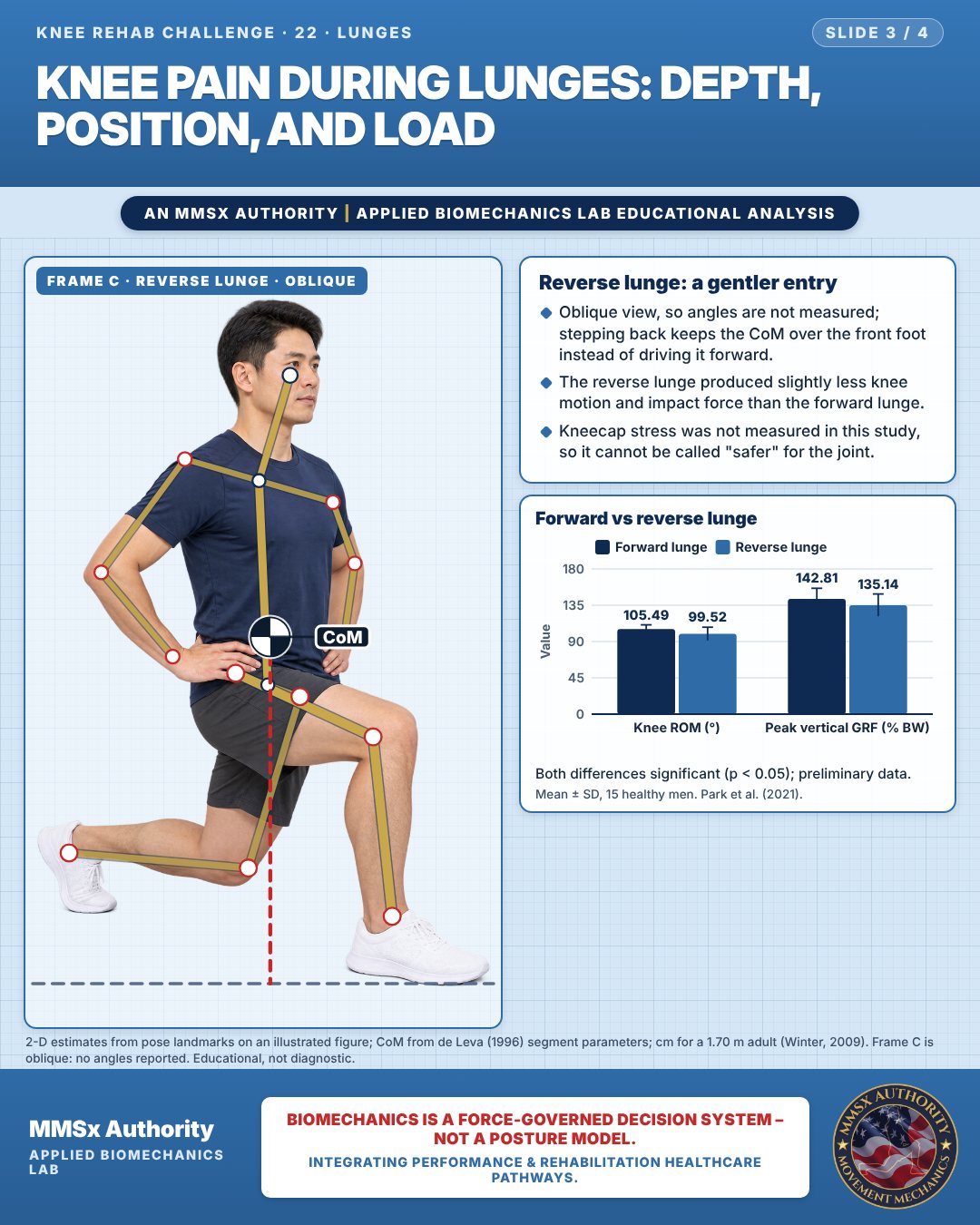
Reverse lunge: a gentler entry
Oblique view, so angles are not measured; stepping back keeps the CoM over the front foot instead of driving it forward.
The reverse lunge produced slightly less knee motion and impact force than the forward lunge.
Kneecap stress was not measured in this study, so it cannot be called "safer" for the joint.
Forward vs reverse lunge
Both differences significant (p < 0.05); preliminary data.
Decision pathway
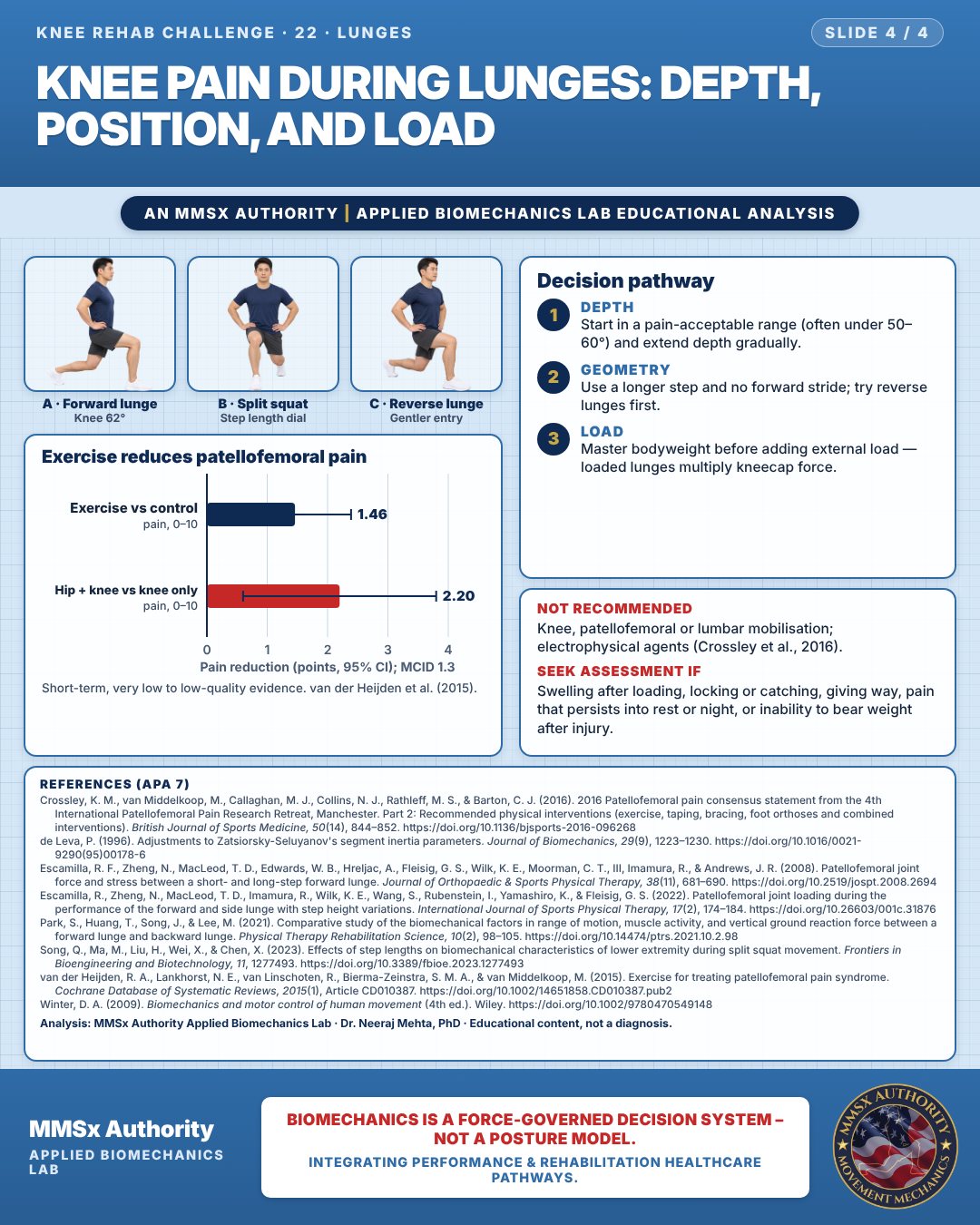
Exercise reduces patellofemoral pain
Depth
Start in a pain-acceptable range (often under 50–60°) and extend depth gradually.
Geometry
Use a longer step and no forward stride; try reverse lunges first.
Load
Master bodyweight before adding external load — loaded lunges multiply kneecap force.
What the evidence supports
- Recommended: exercise therapy; combined hip + knee exercise; combined interventions; foot orthoses short term (Crossley et al., 2016b). Exercise reduces activity pain by a possibly clinically important amount short term (van der Heijden et al., 2015).
- Lunge-specific dose levers with data: limit depth initially (PF stress rises steeply past ~50–60°; Escamilla et al., 2022); longer step / no stride (Escamilla et al., 2008; Song et al., 2023); bodyweight before external load (Escamilla et al., 2022 discussion of 12-RM data); reverse lunge as an option with slightly lower knee excursion and GRF (Park et al., 2021, preliminary).
- Not recommended (PFP consensus): knee/patellofemoral/lumbar mobilisations; electrophysical agents (Crossley et al., 2016b).
Myth and evidence
Myth“Never let the knee go past the toes / never lunge deep — it damages the knee.”
EvidencePF stress rises with knee flexion and with a short step (Escamilla et al., 2008, 2022), so depth and step length are dose dials; peak bodyweight forward-lunge stress (≈1.9 MPa at 80°) is far below 12-RM-loaded values reported as similar to stair ascent/descent (~2,500 N, ~7 MPa — cited in Escamilla et al., 2022). There is no evidence here that lunging per se damages healthy knees; the message is "grade the dose", not "avoid".
Cautions
Not recommended
Knee, patellofemoral or lumbar mobilisation; electrophysical agents (Crossley et al., 2016).
Seek assessment if
Swelling after loading, locking or catching, giving way, pain that persists into rest or night, or inability to bear weight after injury.
Methods note
2-D estimates from pose landmarks on an illustrated figure; CoM from de Leva (1996) segment parameters; cm for a 1.70 m adult (Winter, 2009). Shank angle measured from vertical. Frame C is oblique: no angles reported. These are educational estimates, not a diagnosis.
References
- Crossley, K. M., van Middelkoop, M., Callaghan, M. J., Collins, N. J., Rathleff, M. S., & Barton, C. J. (2016). 2016 Patellofemoral pain consensus statement from the 4th International Patellofemoral Pain Research Retreat, Manchester. Part 2: Recommended physical interventions (exercise, taping, bracing, foot orthoses and combined interventions). British Journal of Sports Medicine, 50(14), 844–852. https://doi.org/10.1136/bjsports-2016-096268
- de Leva, P. (1996). Adjustments to Zatsiorsky-Seluyanov's segment inertia parameters. Journal of Biomechanics, 29(9), 1223–1230. https://doi.org/10.1016/0021-9290(95)00178-6
- Escamilla, R. F., Zheng, N., MacLeod, T. D., Edwards, W. B., Hreljac, A., Fleisig, G. S., Wilk, K. E., Moorman, C. T., III, Imamura, R., & Andrews, J. R. (2008). Patellofemoral joint force and stress between a short- and long-step forward lunge. Journal of Orthopaedic & Sports Physical Therapy, 38(11), 681–690. https://doi.org/10.2519/jospt.2008.2694
- Escamilla, R., Zheng, N., MacLeod, T. D., Imamura, R., Wilk, K. E., Wang, S., Rubenstein, I., Yamashiro, K., & Fleisig, G. S. (2022). Patellofemoral joint loading during the performance of the forward and side lunge with step height variations. International Journal of Sports Physical Therapy, 17(2), 174–184. https://doi.org/10.26603/001c.31876
- Park, S., Huang, T., Song, J., & Lee, M. (2021). Comparative study of the biomechanical factors in range of motion, muscle activity, and vertical ground reaction force between a forward lunge and backward lunge. Physical Therapy Rehabilitation Science, 10(2), 98–105. https://doi.org/10.14474/ptrs.2021.10.2.98
- Song, Q., Ma, M., Liu, H., Wei, X., & Chen, X. (2023). Effects of step lengths on biomechanical characteristics of lower extremity during split squat movement. Frontiers in Bioengineering and Biotechnology, 11, 1277493. https://doi.org/10.3389/fbioe.2023.1277493
- van der Heijden, R. A., Lankhorst, N. E., van Linschoten, R., Bierma-Zeinstra, S. M. A., & van Middelkoop, M. (2015). Exercise for treating patellofemoral pain syndrome. Cochrane Database of Systematic Reviews, 2015(1), Article CD010387. https://doi.org/10.1002/14651858.CD010387.pub2
- Winter, D. A. (2009). Biomechanics and motor control of human movement (4th ed.). Wiley. https://doi.org/10.1002/9780470549148
Cite this article
Mehta, N. (2026, October 23). Knee pain during lunges: depth, position, and load. MMSx Authority Applied Biomechanics Lab. https://mmsxauthority.org/edu/analysis/knee/22-knee-pain-during-lunges-depth-position-and-load/
Part of the collection
Mehta, N. (2026). Knee Rehabilitation: 50 Evidence-Based Biomechanics Analyses [Collection]. MMSx Authority Applied Biomechanics Lab. Zenodo. https://doi.org/10.5281/zenodo.23735752