Knee Rehab Challenge · 21
Knee pain while cycling: looking at position and repetition
Cycling-related knee pain is an overuse complaint — most often anterior knee pain (patellofemoral or patellar/quadriceps tendon region), less often…
Key points
- Check the fit. Set the saddle so the knee is flexed about 25–30° at the bottom of the stroke; avoid a low saddle; change it in small steps.
- Lower the load. Reduce resistance (power) before cadence: knee forces rose with workload, not with pedalling rate.
- Strengthen. For anterior knee pain apply the patellofemoral consensus: hip plus knee exercise (Crossley et al., 2016).
What it is
Cycling-related knee pain is an overuse complaint — most often anterior knee pain (patellofemoral or patellar/quadriceps tendon region), less often medial or lateral (e.g. iliotibial band region) — that develops with repeated pedal cycles; position on the bike (saddle height, set-back, cleat/foot position) and training load (power, volume) both change knee loading.
How we analysed the movement
Three movements relevant to this topic were analysed from illustrated reference figures. Joint centres were located with two-dimensional pose estimation; joint angles, the whole-body centre of mass and, where relevant, the ground-reaction-force line and its moment arm about the knee were then calculated from standard segment parameters. The figures are educational estimates that show how position changes mechanical demand; they are not measurements of a patient. Every research value quoted below comes from the published source cited beside it.
Movement A: Downstroke (side view)
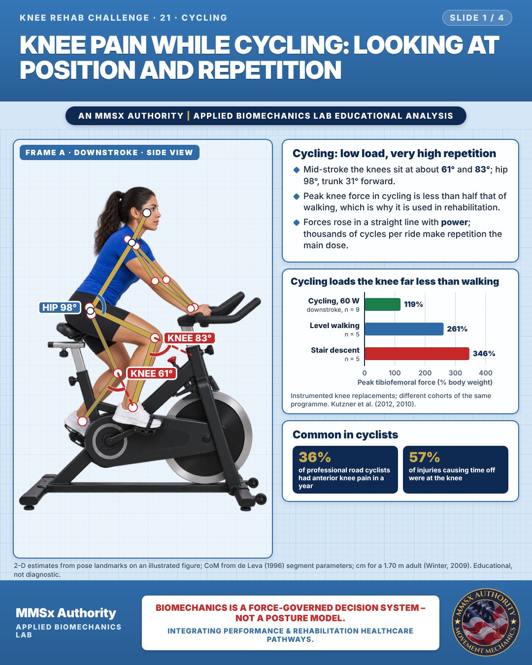
Cycling: low load, very high repetition
Mid-stroke the knees sit at about 61° and 83°; hip 98°, trunk 31° forward.
Peak knee force in cycling is less than half that of walking, which is why it is used in rehabilitation.
Forces rose in a straight line with power; thousands of cycles per ride make repetition the main dose.
Cycling loads the knee far less than walking
Common in cyclists
Movement B: Near top of stroke (side view)
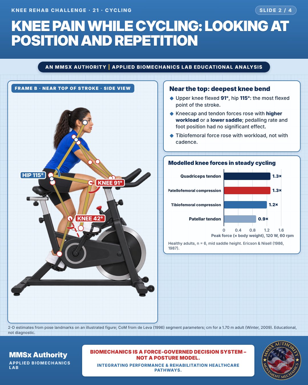
Near the top: deepest knee bend
Upper knee flexed 91°, hip 115°: the most flexed point of the stroke.
Kneecap and tendon forces rose with higher workload or a lower saddle; pedalling rate and foot position had no significant effect.
Tibiofemoral force rose with workload, not with cadence.
Modelled knee forces in steady cycling
Movement C: Knee paths (front view)
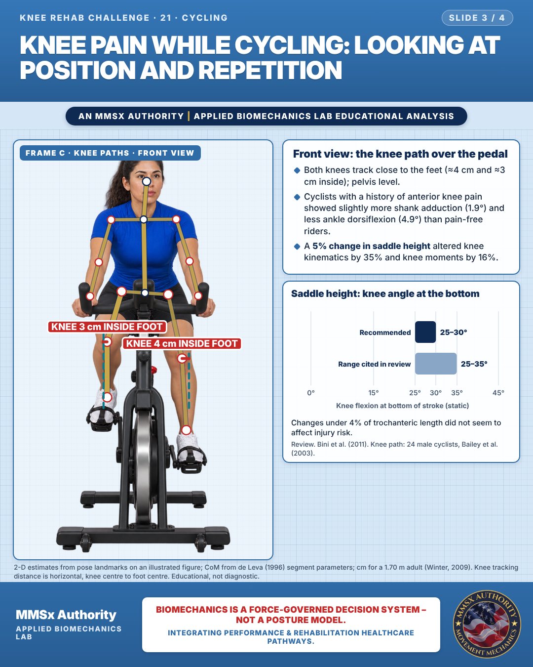
Front view: the knee path over the pedal
Both knees track close to the feet (≈4 cm and ≈3 cm inside); pelvis level.
Cyclists with a history of anterior knee pain showed slightly more shank adduction (1.9°) and less ankle dorsiflexion (4.9°) than pain-free riders.
A 5% change in saddle height altered knee kinematics by 35% and knee moments by 16%.
Saddle height: knee angle at the bottom
Changes under 4% of trochanteric length did not seem to affect injury risk.
Decision pathway
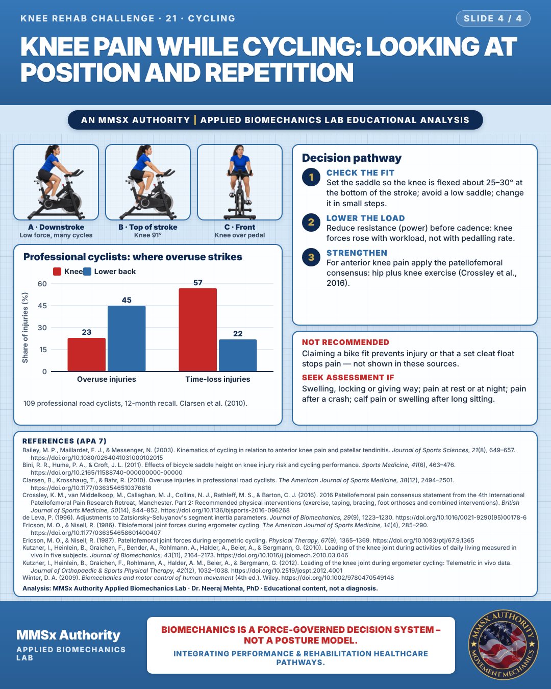
Professional cyclists: where overuse strikes
Check the fit
Set the saddle so the knee is flexed about 25–30° at the bottom of the stroke; avoid a low saddle; change it in small steps.
Lower the load
Reduce resistance (power) before cadence: knee forces rose with workload, not with pedalling rate.
Strengthen
For anterior knee pain apply the patellofemoral consensus: hip plus knee exercise (Crossley et al., 2016).
What the evidence supports
- No cycling-specific clinical practice guideline located. For anterior knee pain in cyclists, apply the patellofemoral pain consensus (exercise therapy with hip + knee focus; combined interventions; foot orthoses short term; not mobilisations or electrophysical agents) (Crossley et al., 2016b).
- Position/dose levers supported by data above: check knee flexion at bottom of stroke (25–30°, Bini et al., 2011); avoid a low saddle (raises PF and tendon forces, Ericson & Nisell, 1987); reduce resistance/power before reducing cadence (forces rise with workload, not with pedalling rate — Ericson & Nisell, 1986, 1987; Kutzner et al., 2012); change saddle in small steps — <4% changes appear not to affect injury risk (Bini et al., 2011).
Myth and evidence
Myth“Pedalling faster (higher cadence) is what hurts the knee.”
EvidenceIn Ericson's laboratory studies pedalling rate had no influence on tibiofemoral or patellofemoral forces, whereas workload and a low saddle raised them (Ericson & Nisell, 1986, 1987). Myth 2: "Cycling loads the knee like walking." In-vivo peak force at 60 W was 119% BW vs 261% BW in walking (Kutzner et al., 2010, 2012).
Cautions
Not recommended
Claiming a bike fit prevents injury or that a set cleat float stops pain — not shown in these sources.
Seek assessment if
Swelling, locking or giving way; pain at rest or at night; pain after a crash; calf pain or swelling after long sitting.
Methods note
2-D estimates from pose landmarks on an illustrated figure; CoM from de Leva (1996) segment parameters; cm for a 1.70 m adult (Winter, 2009). Knee tracking distance is horizontal, knee centre to foot centre. These are educational estimates, not a diagnosis.
References
- Bailey, M. P., Maillardet, F. J., & Messenger, N. (2003). Kinematics of cycling in relation to anterior knee pain and patellar tendinitis. Journal of Sports Sciences, 21(8), 649–657. https://doi.org/10.1080/0264041031000102015
- Bini, R. R., Hume, P. A., & Croft, J. L. (2011). Effects of bicycle saddle height on knee injury risk and cycling performance. Sports Medicine, 41(6), 463–476. https://doi.org/10.2165/11588740-000000000-00000
- Clarsen, B., Krosshaug, T., & Bahr, R. (2010). Overuse injuries in professional road cyclists. The American Journal of Sports Medicine, 38(12), 2494–2501. https://doi.org/10.1177/0363546510376816
- Crossley, K. M., van Middelkoop, M., Callaghan, M. J., Collins, N. J., Rathleff, M. S., & Barton, C. J. (2016). 2016 Patellofemoral pain consensus statement from the 4th International Patellofemoral Pain Research Retreat, Manchester. Part 2: Recommended physical interventions (exercise, taping, bracing, foot orthoses and combined interventions). British Journal of Sports Medicine, 50(14), 844–852. https://doi.org/10.1136/bjsports-2016-096268
- de Leva, P. (1996). Adjustments to Zatsiorsky-Seluyanov's segment inertia parameters. Journal of Biomechanics, 29(9), 1223–1230. https://doi.org/10.1016/0021-9290(95)00178-6
- Ericson, M. O., & Nisell, R. (1986). Tibiofemoral joint forces during ergometer cycling. The American Journal of Sports Medicine, 14(4), 285–290. https://doi.org/10.1177/036354658601400407
- Ericson, M. O., & Nisell, R. (1987). Patellofemoral joint forces during ergometric cycling. Physical Therapy, 67(9), 1365–1369. https://doi.org/10.1093/ptj/67.9.1365
- Kutzner, I., Heinlein, B., Graichen, F., Bender, A., Rohlmann, A., Halder, A., Beier, A., & Bergmann, G. (2010). Loading of the knee joint during activities of daily living measured in vivo in five subjects. Journal of Biomechanics, 43(11), 2164–2173. https://doi.org/10.1016/j.jbiomech.2010.03.046
- Kutzner, I., Heinlein, B., Graichen, F., Rohlmann, A., Halder, A. M., Beier, A., & Bergmann, G. (2012). Loading of the knee joint during ergometer cycling: Telemetric in vivo data. Journal of Orthopaedic & Sports Physical Therapy, 42(12), 1032–1038. https://doi.org/10.2519/jospt.2012.4001
- Winter, D. A. (2009). Biomechanics and motor control of human movement (4th ed.). Wiley. https://doi.org/10.1002/9780470549148
Cite this article
Mehta, N. (2026, October 23). Knee pain while cycling: looking at position and repetition. MMSx Authority Applied Biomechanics Lab. https://mmsxauthority.org/edu/analysis/knee/21-knee-pain-while-cycling-looking-at-position-and-repetition/
Part of the collection
Mehta, N. (2026). Knee Rehabilitation: 50 Evidence-Based Biomechanics Analyses [Collection]. MMSx Authority Applied Biomechanics Lab. Zenodo. https://doi.org/10.5281/zenodo.23735752