Knee Rehab Challenge · 18
Knee pain while walking: what changes under load?
Activity-related knee pain during walking in adults ≥45 years, with no morning stiffness or stiffness ≤30 minutes, is diagnosed clinically as…
Key points
- Keep walking. Exercise and weight management are core (NICE, 2022); pain may rise at first, but regular activity benefits the joint.
- Lower the load. Consider a walking stick in the opposite hand; check footwear. Any weight loss helps; 10% beats 5%.
- Braces. Guidelines differ: ACR/AF strongly recommends a tibiofemoral brace; NICE does not routinely offer braces.
What it is
Activity-related knee pain during walking in adults ≥45 years, with no morning stiffness or stiffness ≤30 minutes, is diagnosed clinically as osteoarthritis without imaging; in younger or atypical cases other diagnoses must be considered.
How we analysed the movement
Three movements relevant to this topic were analysed from illustrated reference figures. Joint centres were located with two-dimensional pose estimation; joint angles, the whole-body centre of mass and, where relevant, the ground-reaction-force line and its moment arm about the knee were then calculated from standard segment parameters. The figures are educational estimates that show how position changes mechanical demand; they are not measurements of a patient. Every research value quoted below comes from the published source cited beside it.
Movement A: Walking, loading (side view)
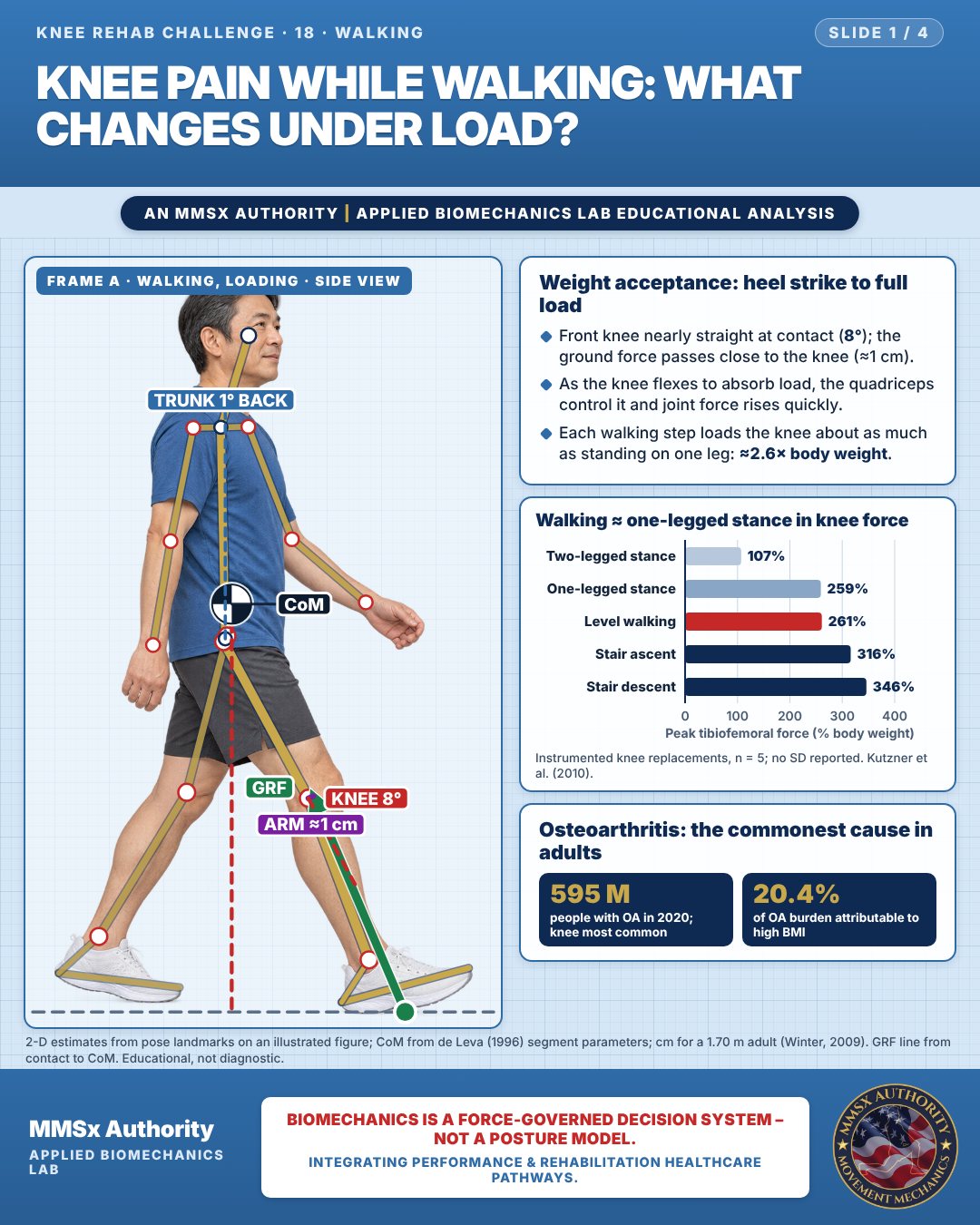
Weight acceptance: heel strike to full load
Front knee nearly straight at contact (8°); the ground force passes close to the knee (≈1 cm).
As the knee flexes to absorb load, the quadriceps control it and joint force rises quickly.
Each walking step loads the knee about as much as standing on one leg: ≈2.6× body weight.
Walking ≈ one-legged stance in knee force
Osteoarthritis: the commonest cause in adults
Movement B: Walking (front view)
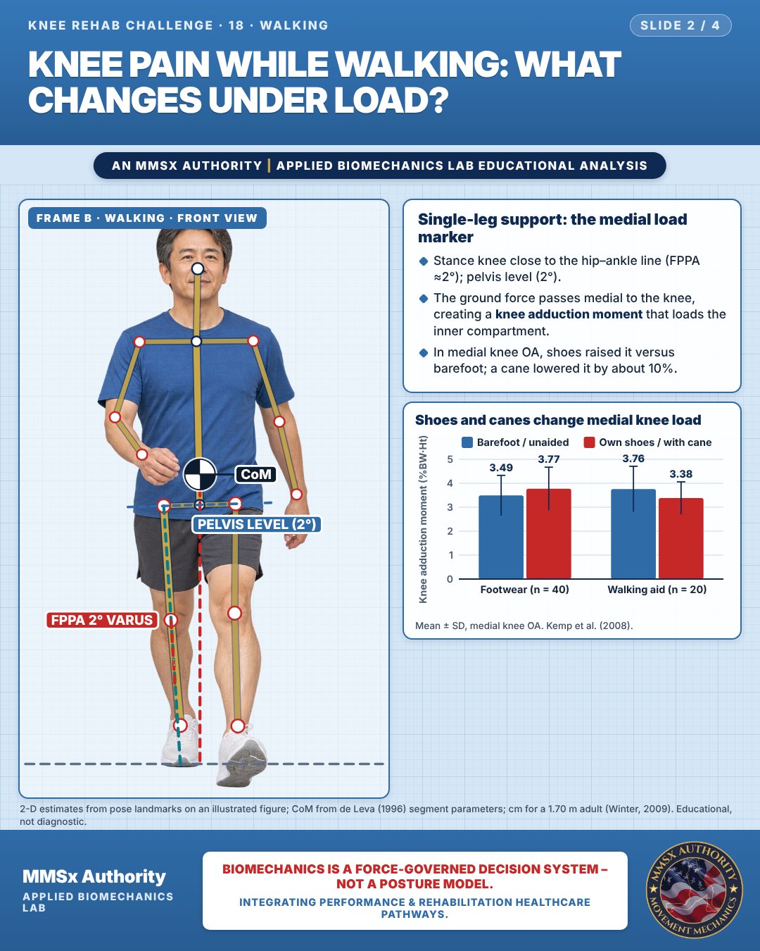
Single-leg support: the medial load marker
Stance knee close to the hip–ankle line (FPPA ≈2°); pelvis level (2°).
The ground force passes medial to the knee, creating a knee adduction moment that loads the inner compartment.
In medial knee OA, shoes raised it versus barefoot; a cane lowered it by about 10%.
Shoes and canes change medial knee load
Movement C: Walking (rear view)
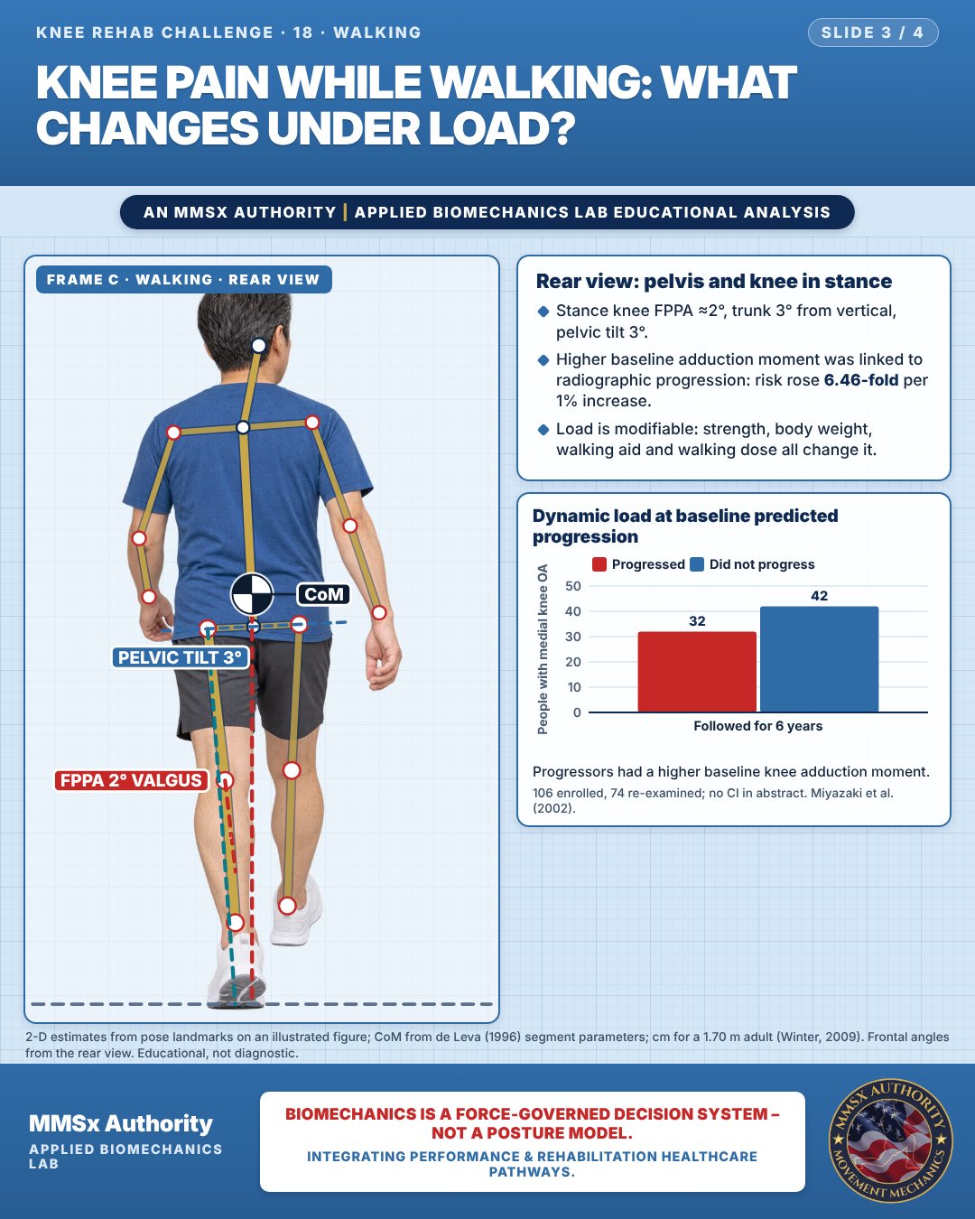
Rear view: pelvis and knee in stance
Stance knee FPPA ≈2°, trunk 3° from vertical, pelvic tilt 3°.
Higher baseline adduction moment was linked to radiographic progression: risk rose 6.46-fold per 1% increase.
Load is modifiable: strength, body weight, walking aid and walking dose all change it.
Dynamic load at baseline predicted progression
Progressors had a higher baseline knee adduction moment.
Decision pathway
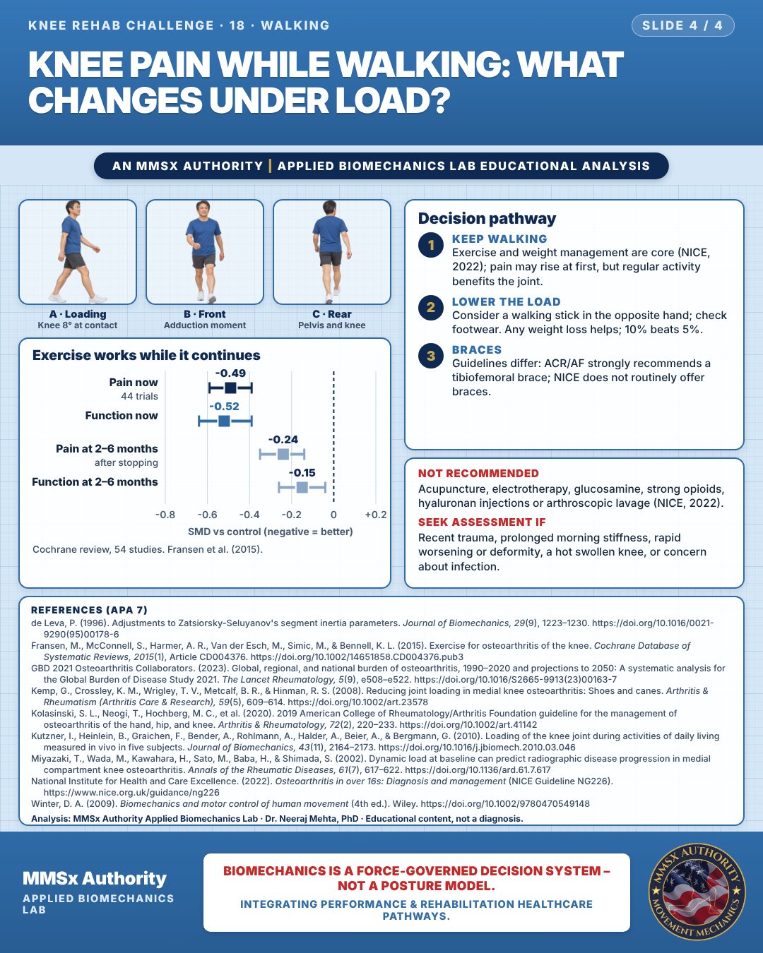
Exercise works while it continues
Keep walking
Exercise and weight management are core (NICE, 2022); pain may rise at first, but regular activity benefits the joint.
Lower the load
Consider a walking stick in the opposite hand; check footwear. Any weight loss helps; 10% beats 5%.
Braces
Guidelines differ: ACR/AF strongly recommends a tibiofemoral brace; NICE does not routinely offer braces.
Cautions
Not recommended
Acupuncture, electrotherapy, glucosamine, strong opioids, hyaluronan injections or arthroscopic lavage (NICE, 2022).
Seek assessment if
Recent trauma, prolonged morning stiffness, rapid worsening or deformity, a hot swollen knee, or concern about infection.
Methods note
2-D estimates from pose landmarks on an illustrated figure; CoM from de Leva (1996) segment parameters; cm for a 1.70 m adult (Winter, 2009). GRF line from contact to CoM. Frontal angles from the rear view. These are educational estimates, not a diagnosis.
References
- de Leva, P. (1996). Adjustments to Zatsiorsky-Seluyanov's segment inertia parameters. Journal of Biomechanics, 29(9), 1223–1230. https://doi.org/10.1016/0021-9290(95)00178-6
- Fransen, M., McConnell, S., Harmer, A. R., Van der Esch, M., Simic, M., & Bennell, K. L. (2015). Exercise for osteoarthritis of the knee. Cochrane Database of Systematic Reviews, 2015(1), Article CD004376. https://doi.org/10.1002/14651858.CD004376.pub3
- GBD 2021 Osteoarthritis Collaborators. (2023). Global, regional, and national burden of osteoarthritis, 1990–2020 and projections to 2050: A systematic analysis for the Global Burden of Disease Study 2021. The Lancet Rheumatology, 5(9), e508–e522. https://doi.org/10.1016/S2665-9913(23)00163-7
- Kemp, G., Crossley, K. M., Wrigley, T. V., Metcalf, B. R., & Hinman, R. S. (2008). Reducing joint loading in medial knee osteoarthritis: Shoes and canes. Arthritis & Rheumatism (Arthritis Care & Research), 59(5), 609–614. https://doi.org/10.1002/art.23578
- Kolasinski, S. L., Neogi, T., Hochberg, M. C., et al. (2020). 2019 American College of Rheumatology/Arthritis Foundation guideline for the management of osteoarthritis of the hand, hip, and knee. Arthritis & Rheumatology, 72(2), 220–233. https://doi.org/10.1002/art.41142
- Kutzner, I., Heinlein, B., Graichen, F., Bender, A., Rohlmann, A., Halder, A., Beier, A., & Bergmann, G. (2010). Loading of the knee joint during activities of daily living measured in vivo in five subjects. Journal of Biomechanics, 43(11), 2164–2173. https://doi.org/10.1016/j.jbiomech.2010.03.046
- Miyazaki, T., Wada, M., Kawahara, H., Sato, M., Baba, H., & Shimada, S. (2002). Dynamic load at baseline can predict radiographic disease progression in medial compartment knee osteoarthritis. Annals of the Rheumatic Diseases, 61(7), 617–622. https://doi.org/10.1136/ard.61.7.617
- National Institute for Health and Care Excellence. (2022). Osteoarthritis in over 16s: Diagnosis and management (NICE Guideline NG226). https://www.nice.org.uk/guidance/ng226
- Winter, D. A. (2009). Biomechanics and motor control of human movement (4th ed.). Wiley. https://doi.org/10.1002/9780470549148
Cite this article
Mehta, N. (2026, October 23). Knee pain while walking: what changes under load? MMSx Authority Applied Biomechanics Lab. https://mmsxauthority.org/edu/analysis/knee/18-knee-pain-while-walking-what-changes-under-load/
Part of the collection
Mehta, N. (2026). Knee Rehabilitation: 50 Evidence-Based Biomechanics Analyses [Collection]. MMSx Authority Applied Biomechanics Lab. Zenodo. https://doi.org/10.5281/zenodo.23735752