Knee Rehab Challenge · 11
MCL injury: when sideways force stresses the knee
A medial collateral ligament (MCL) injury is a sprain or tear of the ligament complex on the inner side of the knee (superficial MCL, deep MCL,…
Key points
- Grade it. Examine for laxity at 0° and 30° of bend; laxity in full extension suggests the posteromedial corner is involved.
- Rehabilitate. Grade I–II: weight-bear as tolerated, progressive range, strength, running and agility; return usually in 3–12 weeks.
- Escalate. Combined ACL + MCL injuries need specialist care: untreated MCL laxity raised ACL graft revision risk.
What it is
A medial collateral ligament (MCL) injury is a sprain or tear of the ligament complex on the inner side of the knee (superficial MCL, deep MCL, posterior oblique ligament), usually caused by a valgus (knee-inward) force — most often a blow to the outside of the knee — and graded I (stretch), II (partial tear) or III (complete tear) (Lundblad et al., 2019; Ball et al., 2020).
How we analysed the movement
Three movements relevant to this topic were analysed from illustrated reference figures. Joint centres were located with two-dimensional pose estimation; joint angles, the whole-body centre of mass and, where relevant, the ground-reaction-force line and its moment arm about the knee were then calculated from standard segment parameters. The figures are educational estimates that show how position changes mechanical demand; they are not measurements of a patient. Every research value quoted below comes from the published source cited beside it.
Movement A: Side contact (front view)
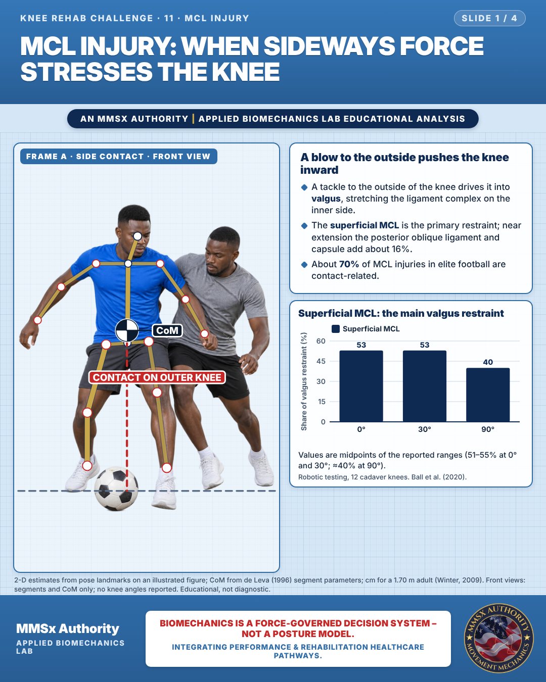
A blow to the outside pushes the knee inward
A tackle to the outside of the knee drives it into valgus, stretching the ligament complex on the inner side.
The superficial MCL is the primary restraint; near extension the posterior oblique ligament and capsule add about 16%.
About 70% of MCL injuries in elite football are contact-related.
Superficial MCL: the main valgus restraint
Values are midpoints of the reported ranges (51–55% at 0° and 30°; ≈40% at 90°).
Movement B: Planted lateral move (front view)
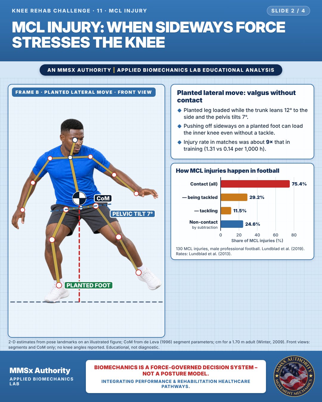
Planted lateral move: valgus without contact
Planted leg loaded while the trunk leans 12° to the side and the pelvis tilts 7°.
Pushing off sideways on a planted foot can load the inner knee even without a tackle.
Injury rate in matches was about 9× that in training (1.31 vs 0.14 per 1,000 h).
How MCL injuries happen in football
Movement C: Seated after the incident (front view)
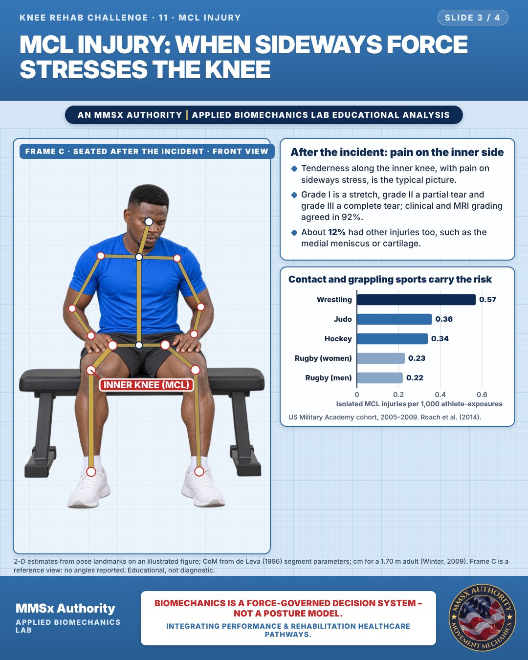
After the incident: pain on the inner side
Tenderness along the inner knee, with pain on sideways stress, is the typical picture.
Grade I is a stretch, grade II a partial tear and grade III a complete tear; clinical and MRI grading agreed in 92%.
About 12% had other injuries too, such as the medial meniscus or cartilage.
Contact and grappling sports carry the risk
Decision pathway
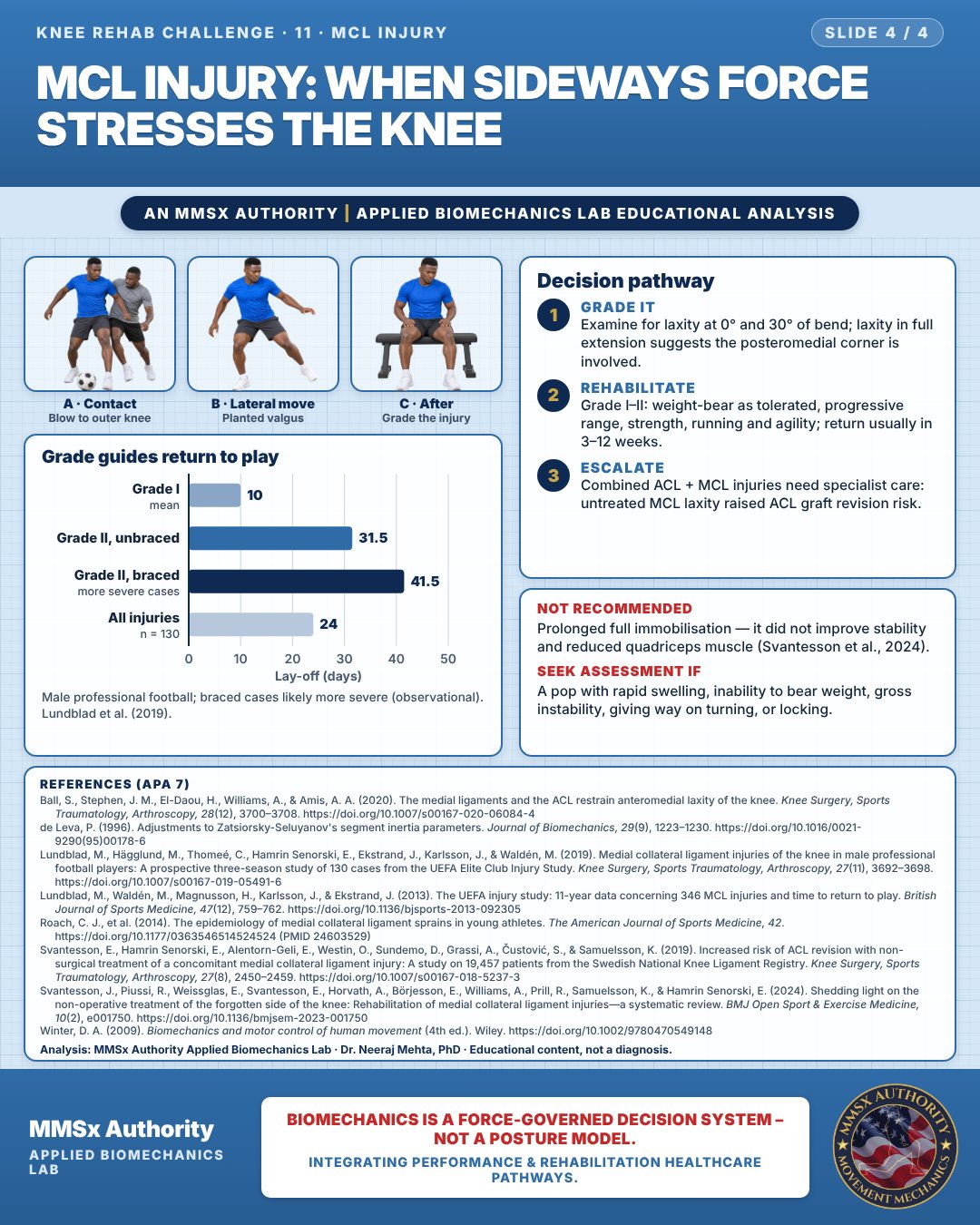
Grade guides return to play
Grade it
Examine for laxity at 0° and 30° of bend; laxity in full extension suggests the posteromedial corner is involved.
Rehabilitate
Grade I–II: weight-bear as tolerated, progressive range, strength, running and agility; return usually in 3–12 weeks.
Escalate
Combined ACL + MCL injuries need specialist care: untreated MCL laxity raised ACL graft revision risk.
What the evidence supports
- Note on bracing: no direct brace vs no-brace comparison exists; in professional football, braced grade II injuries had a longer lay-off (observational, confounded by severity) (Lundblad et al., 2019; Svantesson et al., 2024).
- Combined ACL + MCL injuries need specialist assessment: residual MCL laxity left untreated is associated with higher ACL graft revision risk (Svantesson et al., 2019).
- Not recommended: prolonged full immobilisation — complete immobilisation did not improve valgus stability at 2 years and reduced quadriceps muscle fibre number/size (Svantesson et al., 2024).
Myth and evidence
Myth“MCL tears always need a brace and surgery.”
EvidenceMost isolated MCL injuries are treated non-operatively with return in ~3–12 weeks (Svantesson et al., 2024); grade I mean lay-off ~10 days in elite football (Lundblad et al., 2019).
Myth“MCL injuries are mostly non-contact twisting injuries.”
Evidence~70–75% are contact-related in professional football (Lundblad et al., 2013; 2019).
Cautions
Not recommended
Prolonged full immobilisation — it did not improve stability and reduced quadriceps muscle (Svantesson et al., 2024).
Seek assessment if
A pop with rapid swelling, inability to bear weight, gross instability, giving way on turning, or locking.
Methods note
2-D estimates from pose landmarks on an illustrated figure; CoM from de Leva (1996) segment parameters; cm for a 1.70 m adult (Winter, 2009). Front views: segments and CoM only; no knee angles reported. Frame C is a reference view: no angles reported. These are educational estimates, not a diagnosis.
References
- Ball, S., Stephen, J. M., El-Daou, H., Williams, A., & Amis, A. A. (2020). The medial ligaments and the ACL restrain anteromedial laxity of the knee. Knee Surgery, Sports Traumatology, Arthroscopy, 28(12), 3700–3708. https://doi.org/10.1007/s00167-020-06084-4
- de Leva, P. (1996). Adjustments to Zatsiorsky-Seluyanov's segment inertia parameters. Journal of Biomechanics, 29(9), 1223–1230. https://doi.org/10.1016/0021-9290(95)00178-6
- Lundblad, M., Hägglund, M., Thomeé, C., Hamrin Senorski, E., Ekstrand, J., Karlsson, J., & Waldén, M. (2019). Medial collateral ligament injuries of the knee in male professional football players: A prospective three-season study of 130 cases from the UEFA Elite Club Injury Study. Knee Surgery, Sports Traumatology, Arthroscopy, 27(11), 3692–3698. https://doi.org/10.1007/s00167-019-05491-6
- Lundblad, M., Waldén, M., Magnusson, H., Karlsson, J., & Ekstrand, J. (2013). The UEFA injury study: 11-year data concerning 346 MCL injuries and time to return to play. British Journal of Sports Medicine, 47(12), 759–762. https://doi.org/10.1136/bjsports-2013-092305
- Roach, C. J., et al. (2014). The epidemiology of medial collateral ligament sprains in young athletes. The American Journal of Sports Medicine, 42. https://doi.org/10.1177/0363546514524524 (PMID 24603529)
- Svantesson, E., Hamrin Senorski, E., Alentorn-Geli, E., Westin, O., Sundemo, D., Grassi, A., Čustović, S., & Samuelsson, K. (2019). Increased risk of ACL revision with non-surgical treatment of a concomitant medial collateral ligament injury: A study on 19,457 patients from the Swedish National Knee Ligament Registry. Knee Surgery, Sports Traumatology, Arthroscopy, 27(8), 2450–2459. https://doi.org/10.1007/s00167-018-5237-3
- Svantesson, J., Piussi, R., Weissglas, E., Svantesson, E., Horvath, A., Börjesson, E., Williams, A., Prill, R., Samuelsson, K., & Hamrin Senorski, E. (2024). Shedding light on the non-operative treatment of the forgotten side of the knee: Rehabilitation of medial collateral ligament injuries—a systematic review. BMJ Open Sport & Exercise Medicine, 10(2), e001750. https://doi.org/10.1136/bmjsem-2023-001750
- Winter, D. A. (2009). Biomechanics and motor control of human movement (4th ed.). Wiley. https://doi.org/10.1002/9780470549148
Cite this article
Mehta, N. (2026, October 23). MCL injury: when sideways force stresses the knee. MMSx Authority Applied Biomechanics Lab. https://mmsxauthority.org/edu/analysis/knee/11-mcl-injury-when-sideways-force-stresses-the-knee/
Part of the collection
Mehta, N. (2026). Knee Rehabilitation: 50 Evidence-Based Biomechanics Analyses [Collection]. MMSx Authority Applied Biomechanics Lab. Zenodo. https://doi.org/10.5281/zenodo.23735752