Knee Rehab Challenge · 10
Kneecap instability: when the kneecap feels like it slips
Lateral patellar instability is the tendency of the kneecap to sublux or dislocate out of the trochlear groove towards the outside of the knee,…
Key points
- Assess risk. Check for trochlear dysplasia, patella alta, age under 18 and TT–TG distance: they drive recurrence.
- Rehabilitate. Quadriceps (including VMO) and hip control, then graded return to turning and cutting.
- Consider surgery. Recurrent instability or high-risk anatomy: discuss stabilisation such as MPFL reconstruction.
What it is
Lateral patellar instability is the tendency of the kneecap to sublux or dislocate out of the trochlear groove towards the outside of the knee, typically during twisting on a planted foot with the knee near extension; it is driven by an interaction of soft-tissue restraint (chiefly the medial patellofemoral ligament, MPFL) and bony anatomy (trochlear shape, patellar height, tubercle position) (Desio et al., 1998; Christensen et al., 2017).
How we analysed the movement
Three movements relevant to this topic were analysed from illustrated reference figures. Joint centres were located with two-dimensional pose estimation; joint angles, the whole-body centre of mass and, where relevant, the ground-reaction-force line and its moment arm about the knee were then calculated from standard segment parameters. The figures are educational estimates that show how position changes mechanical demand; they are not measurements of a patient. Every research value quoted below comes from the published source cited beside it.
Movement A: Planted-foot turn, oblique
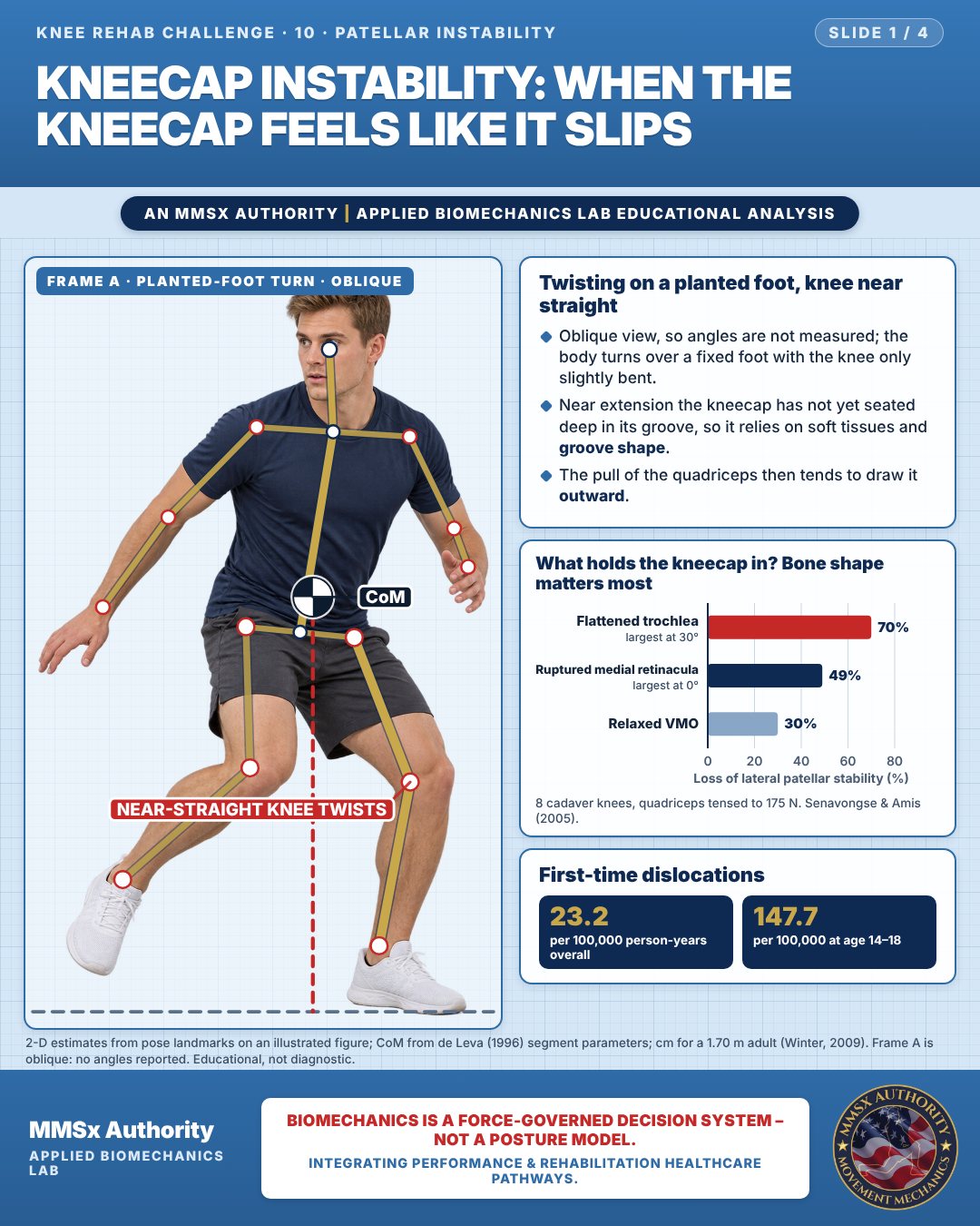
Twisting on a planted foot, knee near straight
Oblique view, so angles are not measured; the body turns over a fixed foot with the knee only slightly bent.
Near extension the kneecap has not yet seated deep in its groove, so it relies on soft tissues and groove shape.
The pull of the quadriceps then tends to draw it outward.
What holds the kneecap in? Bone shape matters most
First-time dislocations
Movement B: Dance turn, oblique
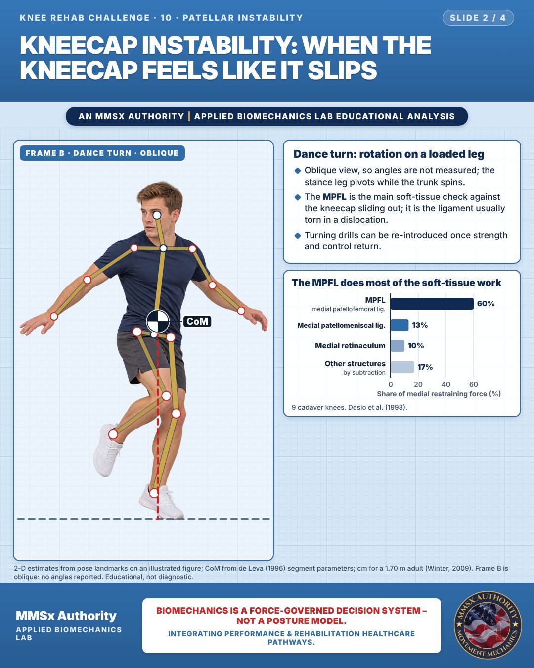
Dance turn: rotation on a loaded leg
Oblique view, so angles are not measured; the stance leg pivots while the trunk spins.
The MPFL is the main soft-tissue check against the kneecap sliding out; it is the ligament usually torn in a dislocation.
Turning drills can be re-introduced once strength and control return.
The MPFL does most of the soft-tissue work
Movement C: Seated after an episode, oblique
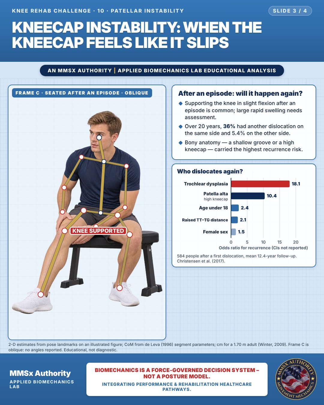
After an episode: will it happen again?
Supporting the knee in slight flexion after an episode is common; large rapid swelling needs assessment.
Over 20 years, 36% had another dislocation on the same side and 5.4% on the other side.
Bony anatomy — a shallow groove or a high kneecap — carried the highest recurrence risk.
Who dislocates again?
Decision pathway
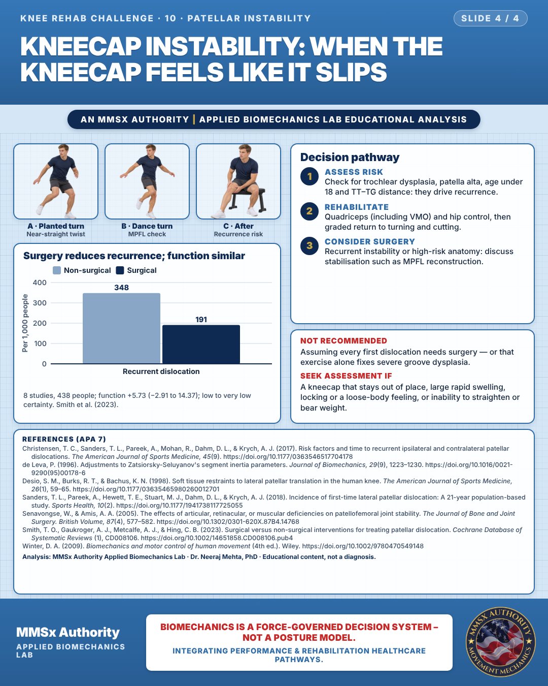
Surgery reduces recurrence; function similar
Assess risk
Check for trochlear dysplasia, patella alta, age under 18 and TT–TG distance: they drive recurrence.
Rehabilitate
Quadriceps (including VMO) and hip control, then graded return to turning and cutting.
Consider surgery
Recurrent instability or high-risk anatomy: discuss stabilisation such as MPFL reconstruction.
What the evidence supports
- Evidence: primary surgical management reduces recurrence compared with non-surgical care but does not clearly improve function; certainty is low to very low (Smith et al., 2023). Treatment choice should therefore weigh individual recurrence risk — trochlear dysplasia, patella alta, age < 18, raised TT–TG (Christensen et al., 2017).
Myth and evidence
Myth“A weak VMO is the main reason kneecaps slip.”
EvidenceIn vitro, a flattened trochlea reduced lateral stability by 70% vs 30% for VMO relaxation (Senavongse & Amis, 2005); trochlear dysplasia carried OR 18.1 for recurrence (Christensen et al., 2017). Muscle matters, but bone shape matters more.
Myth“Once it goes back in, it is fixed.”
Evidence36% cumulative ipsilateral recurrence at 20 years (Christensen et al., 2017).
Cautions
Not recommended
Assuming every first dislocation needs surgery — or that exercise alone fixes severe groove dysplasia.
Seek assessment if
A kneecap that stays out of place, large rapid swelling, locking or a loose-body feeling, or inability to straighten or bear weight.
Methods note
2-D estimates from pose landmarks on an illustrated figure; CoM from de Leva (1996) segment parameters; cm for a 1.70 m adult (Winter, 2009). Frame A is oblique: no angles reported. Frame B is oblique: no angles reported. Frame C is oblique: no angles reported. These are educational estimates, not a diagnosis.
References
- Christensen, T. C., Sanders, T. L., Pareek, A., Mohan, R., Dahm, D. L., & Krych, A. J. (2017). Risk factors and time to recurrent ipsilateral and contralateral patellar dislocations. The American Journal of Sports Medicine, 45(9). https://doi.org/10.1177/0363546517704178
- de Leva, P. (1996). Adjustments to Zatsiorsky-Seluyanov's segment inertia parameters. Journal of Biomechanics, 29(9), 1223–1230. https://doi.org/10.1016/0021-9290(95)00178-6
- Desio, S. M., Burks, R. T., & Bachus, K. N. (1998). Soft tissue restraints to lateral patellar translation in the human knee. The American Journal of Sports Medicine, 26(1), 59–65. https://doi.org/10.1177/03635465980260012701
- Sanders, T. L., Pareek, A., Hewett, T. E., Stuart, M. J., Dahm, D. L., & Krych, A. J. (2018). Incidence of first-time lateral patellar dislocation: A 21-year population-based study. Sports Health, 10(2). https://doi.org/10.1177/1941738117725055
- Senavongse, W., & Amis, A. A. (2005). The effects of articular, retinacular, or muscular deficiencies on patellofemoral joint stability. The Journal of Bone and Joint Surgery. British Volume, 87(4), 577–582. https://doi.org/10.1302/0301-620X.87B4.14768
- Smith, T. O., Gaukroger, A. J., Metcalfe, A. J., & Hing, C. B. (2023). Surgical versus non-surgical interventions for treating patellar dislocation. Cochrane Database of Systematic Reviews (1), CD008106. https://doi.org/10.1002/14651858.CD008106.pub4
- Winter, D. A. (2009). Biomechanics and motor control of human movement (4th ed.). Wiley. https://doi.org/10.1002/9780470549148
Cite this article
Mehta, N. (2026, October 23). Kneecap instability: when the kneecap feels like it slips. MMSx Authority Applied Biomechanics Lab. https://mmsxauthority.org/edu/analysis/knee/10-kneecap-instability-when-the-kneecap-feels-like-it-slips/
Part of the collection
Mehta, N. (2026). Knee Rehabilitation: 50 Evidence-Based Biomechanics Analyses [Collection]. MMSx Authority Applied Biomechanics Lab. Zenodo. https://doi.org/10.5281/zenodo.23735752