Knee Rehab Challenge · 09
Knee pain when kneeling: understanding prepatellar bursitis
Prepatellar bursitis is inflammation and swelling of the superficial bursa lying between the skin and the front of the kneecap.
Key points
- Protect. Reduce kneeling time, use kneepads or a cushion, and kneel near 90° rather than on one knee; never strike with the knee.
- Settle. Non-infected bursitis: rest from pressure, ice, compression, anti-inflammatories; aspiration if large.
- Treat infection. Suspected infection needs aspiration and antibiotics; steroid injection only once infection is excluded.
What it is
Prepatellar bursitis is inflammation and swelling of the superficial bursa lying between the skin and the front of the kneecap; it is most often non-infective (repetitive pressure/friction or a blow) but about one in three cases is septic (infected), usually via a skin breach (Baumbach et al., 2014).
How we analysed the movement
Three movements relevant to this topic were analysed from illustrated reference figures. Joint centres were located with two-dimensional pose estimation; joint angles, the whole-body centre of mass and, where relevant, the ground-reaction-force line and its moment arm about the knee were then calculated from standard segment parameters. The figures are educational estimates that show how position changes mechanical demand; they are not measurements of a patient. Every research value quoted below comes from the published source cited beside it.
Movement A: Kneeling gardening (side view)
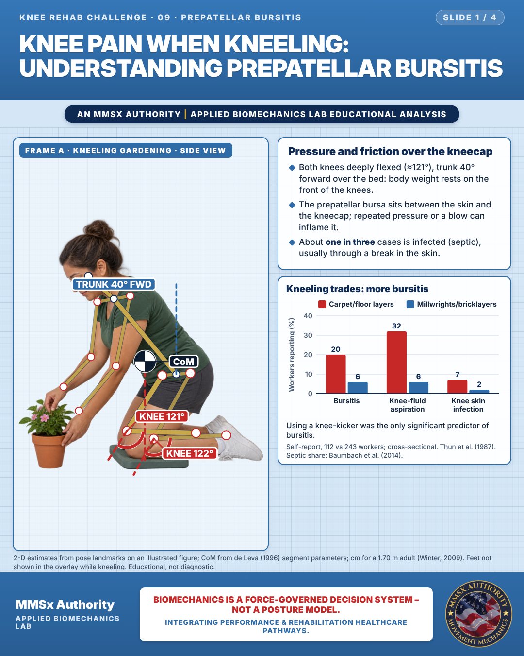
Pressure and friction over the kneecap
Both knees deeply flexed (≈121°), trunk 40° forward over the bed: body weight rests on the front of the knees.
The prepatellar bursa sits between the skin and the kneecap; repeated pressure or a blow can inflame it.
About one in three cases is infected (septic), usually through a break in the skin.
Kneeling trades: more bursitis
Using a knee-kicker was the only significant predictor of bursitis.
Movement B: Kneeling at a low shelf (side view)
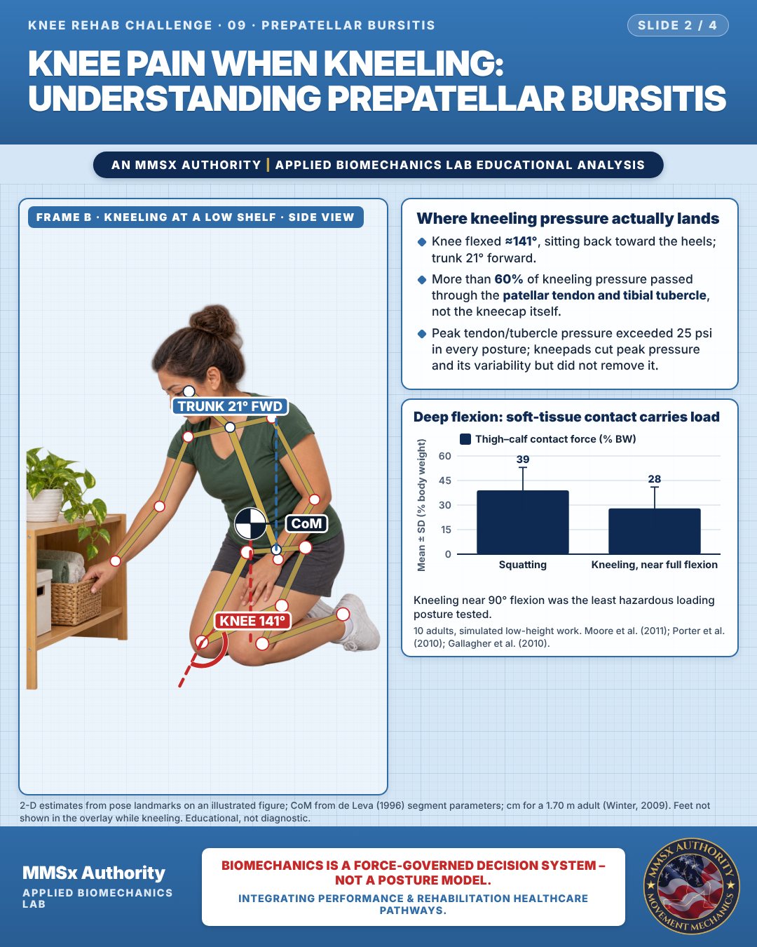
Where kneeling pressure actually lands
Knee flexed ≈141°, sitting back toward the heels; trunk 21° forward.
More than 60% of kneeling pressure passed through the patellar tendon and tibial tubercle, not the kneecap itself.
Peak tendon/tubercle pressure exceeded 25 psi in every posture; kneepads cut peak pressure and its variability but did not remove it.
Deep flexion: soft-tissue contact carries load
Kneeling near 90° flexion was the least hazardous loading posture tested.
Movement C: Half-kneeling rise (side view)
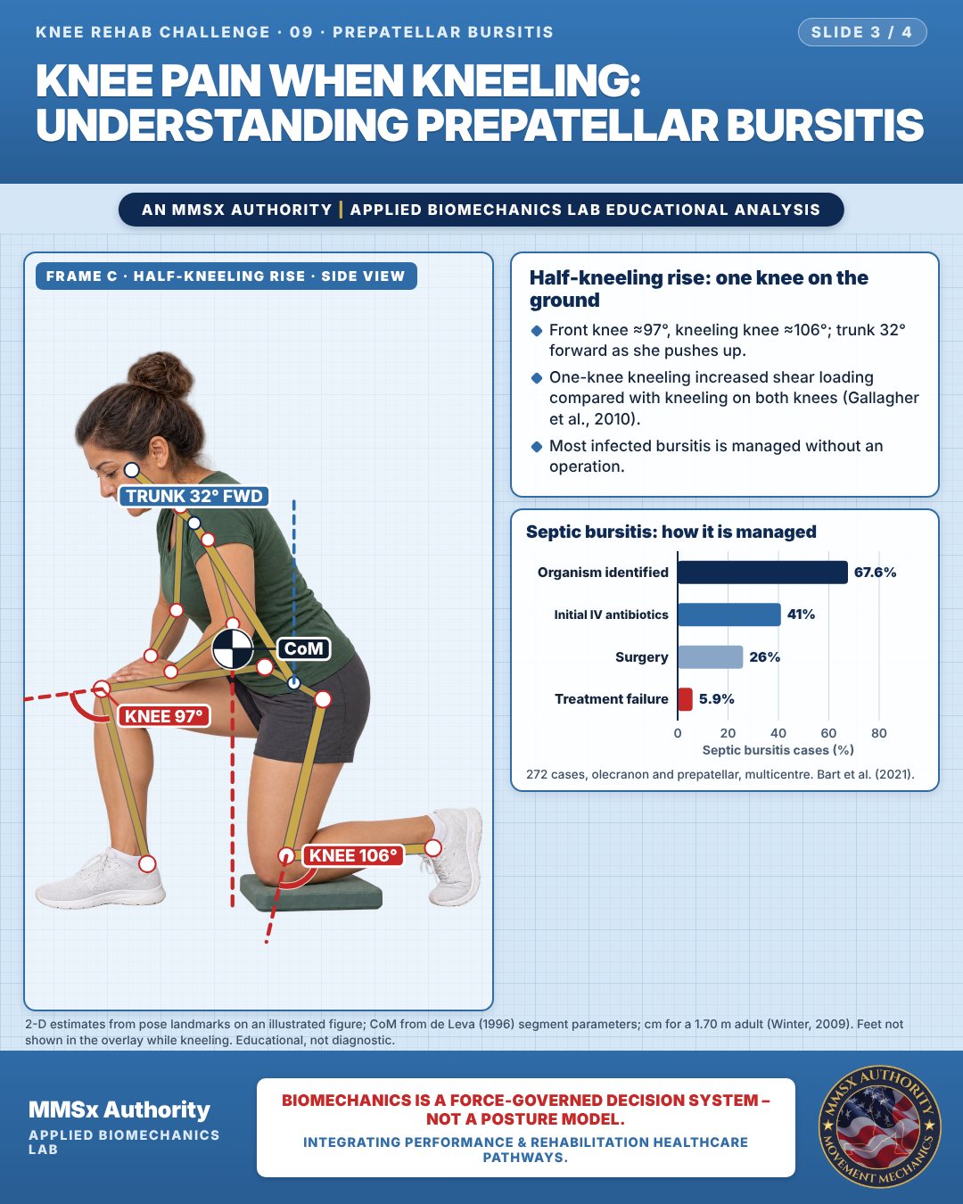
Half-kneeling rise: one knee on the ground
Front knee ≈97°, kneeling knee ≈106°; trunk 32° forward as she pushes up.
One-knee kneeling increased shear loading compared with kneeling on both knees (Gallagher et al., 2010).
Most infected bursitis is managed without an operation.
Septic bursitis: how it is managed
Decision pathway
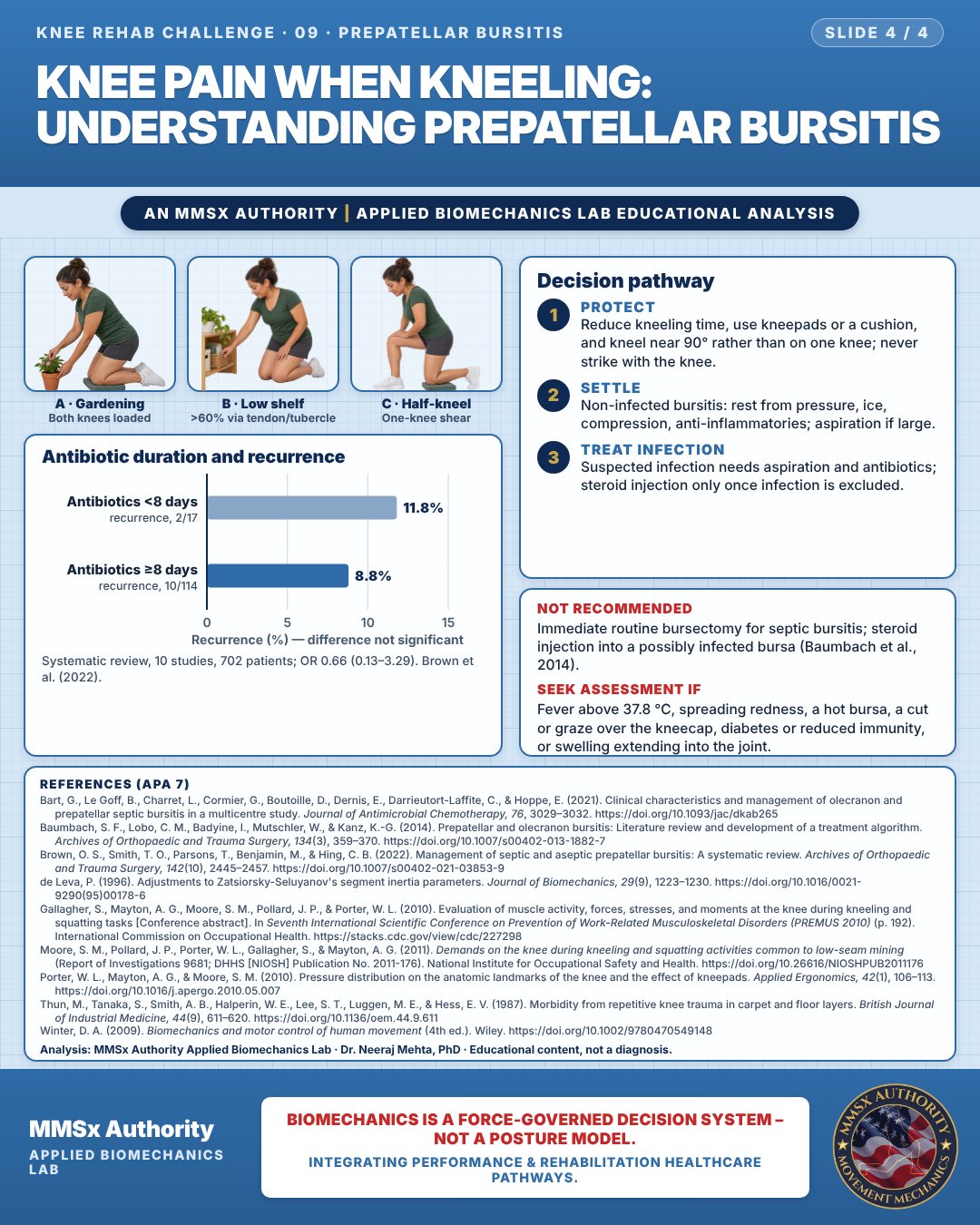
Antibiotic duration and recurrence
Protect
Reduce kneeling time, use kneepads or a cushion, and kneel near 90° rather than on one knee; never strike with the knee.
Settle
Non-infected bursitis: rest from pressure, ice, compression, anti-inflammatories; aspiration if large.
Treat infection
Suspected infection needs aspiration and antibiotics; steroid injection only once infection is excluded.
What the evidence supports
- Not recommended: routine immediate bursectomy for septic bursitis — "the available evidence did not support" it (Baumbach et al., 2014). Steroid injection into a possibly infected bursa.
- Prevention: reduce kneeling time/pressure; kneepads reduce peak pressure over the patellar tendon/tibial tubercle but do not remove it (Porter et al., 2010); avoid striking with the knee (knee-kicker use was the predictor in Thun et al., 1987).
Myth and evidence
Myth“Kneeling puts all the pressure on the kneecap.”
Evidence> 60% of kneeling pressure passes through the patellar tendon/tibial tubercle; patella mean pressures were < 10 psi, and < 3 psi near full flexion (Porter et al., 2010).
Myth“An infected bursa always needs an operation.”
EvidenceSurgery used in 26% of septic cases with 5.9% overall failure (Bart et al., 2021); review evidence does not support immediate bursectomy (Baumbach et al., 2014).
Cautions
Not recommended
Immediate routine bursectomy for septic bursitis; steroid injection into a possibly infected bursa (Baumbach et al., 2014).
Seek assessment if
Fever above 37.8 °C, spreading redness, a hot bursa, a cut or graze over the kneecap, diabetes or reduced immunity, or swelling extending into the joint.
Methods note
2-D estimates from pose landmarks on an illustrated figure; CoM from de Leva (1996) segment parameters; cm for a 1.70 m adult (Winter, 2009). Feet not shown in the overlay while kneeling. These are educational estimates, not a diagnosis.
References
- Bart, G., Le Goff, B., Charret, L., Cormier, G., Boutoille, D., Dernis, E., Darrieutort-Laffite, C., & Hoppe, E. (2021). Clinical characteristics and management of olecranon and prepatellar septic bursitis in a multicentre study. Journal of Antimicrobial Chemotherapy, 76, 3029–3032. https://doi.org/10.1093/jac/dkab265
- Baumbach, S. F., Lobo, C. M., Badyine, I., Mutschler, W., & Kanz, K.-G. (2014). Prepatellar and olecranon bursitis: Literature review and development of a treatment algorithm. Archives of Orthopaedic and Trauma Surgery, 134(3), 359–370. https://doi.org/10.1007/s00402-013-1882-7
- Brown, O. S., Smith, T. O., Parsons, T., Benjamin, M., & Hing, C. B. (2022). Management of septic and aseptic prepatellar bursitis: A systematic review. Archives of Orthopaedic and Trauma Surgery, 142(10), 2445–2457. https://doi.org/10.1007/s00402-021-03853-9
- de Leva, P. (1996). Adjustments to Zatsiorsky-Seluyanov's segment inertia parameters. Journal of Biomechanics, 29(9), 1223–1230. https://doi.org/10.1016/0021-9290(95)00178-6
- Gallagher, S., Mayton, A. G., Moore, S. M., Pollard, J. P., & Porter, W. L. (2010). Evaluation of muscle activity, forces, stresses, and moments at the knee during kneeling and squatting tasks [Conference abstract]. In Seventh International Scientific Conference on Prevention of Work-Related Musculoskeletal Disorders (PREMUS 2010) (p. 192). International Commission on Occupational Health. https://stacks.cdc.gov/view/cdc/227298
- Moore, S. M., Pollard, J. P., Porter, W. L., Gallagher, S., & Mayton, A. G. (2011). Demands on the knee during kneeling and squatting activities common to low-seam mining (Report of Investigations 9681; DHHS [NIOSH] Publication No. 2011-176). National Institute for Occupational Safety and Health. https://doi.org/10.26616/NIOSHPUB2011176
- Porter, W. L., Mayton, A. G., & Moore, S. M. (2010). Pressure distribution on the anatomic landmarks of the knee and the effect of kneepads. Applied Ergonomics, 42(1), 106–113. https://doi.org/10.1016/j.apergo.2010.05.007
- Thun, M., Tanaka, S., Smith, A. B., Halperin, W. E., Lee, S. T., Luggen, M. E., & Hess, E. V. (1987). Morbidity from repetitive knee trauma in carpet and floor layers. British Journal of Industrial Medicine, 44(9), 611–620. https://doi.org/10.1136/oem.44.9.611
- Winter, D. A. (2009). Biomechanics and motor control of human movement (4th ed.). Wiley. https://doi.org/10.1002/9780470549148
Cite this article
Mehta, N. (2026, October 23). Knee pain when kneeling: understanding prepatellar bursitis. MMSx Authority Applied Biomechanics Lab. https://mmsxauthority.org/edu/analysis/knee/09-knee-pain-when-kneeling-understanding-prepatellar-bursitis/
Part of the collection
Mehta, N. (2026). Knee Rehabilitation: 50 Evidence-Based Biomechanics Analyses [Collection]. MMSx Authority Applied Biomechanics Lab. Zenodo. https://doi.org/10.5281/zenodo.23735752