Knee Rehab Challenge · 08
Pain behind the knee: could it be a Baker's cyst?
A popliteal (Baker's) cyst is a fluid-filled distension of the gastrocnemius–semimembranosus bursa at the back of the knee, which in adults usually…
Key points
- Screen. Calf pain, swelling, redness or warmth needs an ultrasound: a ruptured cyst can mimic a deep-vein clot, and the two can coexist.
- Treat the knee. At least 6 weeks of non-operative care keeping knee movement; a joint injection can shrink the cyst. Manage the effusion, meniscus or OA behind it.
- Escalate. Persistent cases: arthroscopy for the joint cause first; excision only for large, highly symptomatic cysts or nerve/vessel compression.
What it is
A popliteal (Baker's) cyst is a fluid-filled distension of the gastrocnemius–semimembranosus bursa at the back of the knee, which in adults usually communicates with the knee joint and is usually secondary to an intra-articular problem (effusion, meniscal tear, osteoarthritis) (Frush & Noyes, 2015; Sansone et al., 1995).
How we analysed the movement
Three movements relevant to this topic were analysed from illustrated reference figures. Joint centres were located with two-dimensional pose estimation; joint angles, the whole-body centre of mass and, where relevant, the ground-reaction-force line and its moment arm about the knee were then calculated from standard segment parameters. The figures are educational estimates that show how position changes mechanical demand; they are not measurements of a patient. Every research value quoted below comes from the published source cited beside it.
Movement A: Standing (rear view)
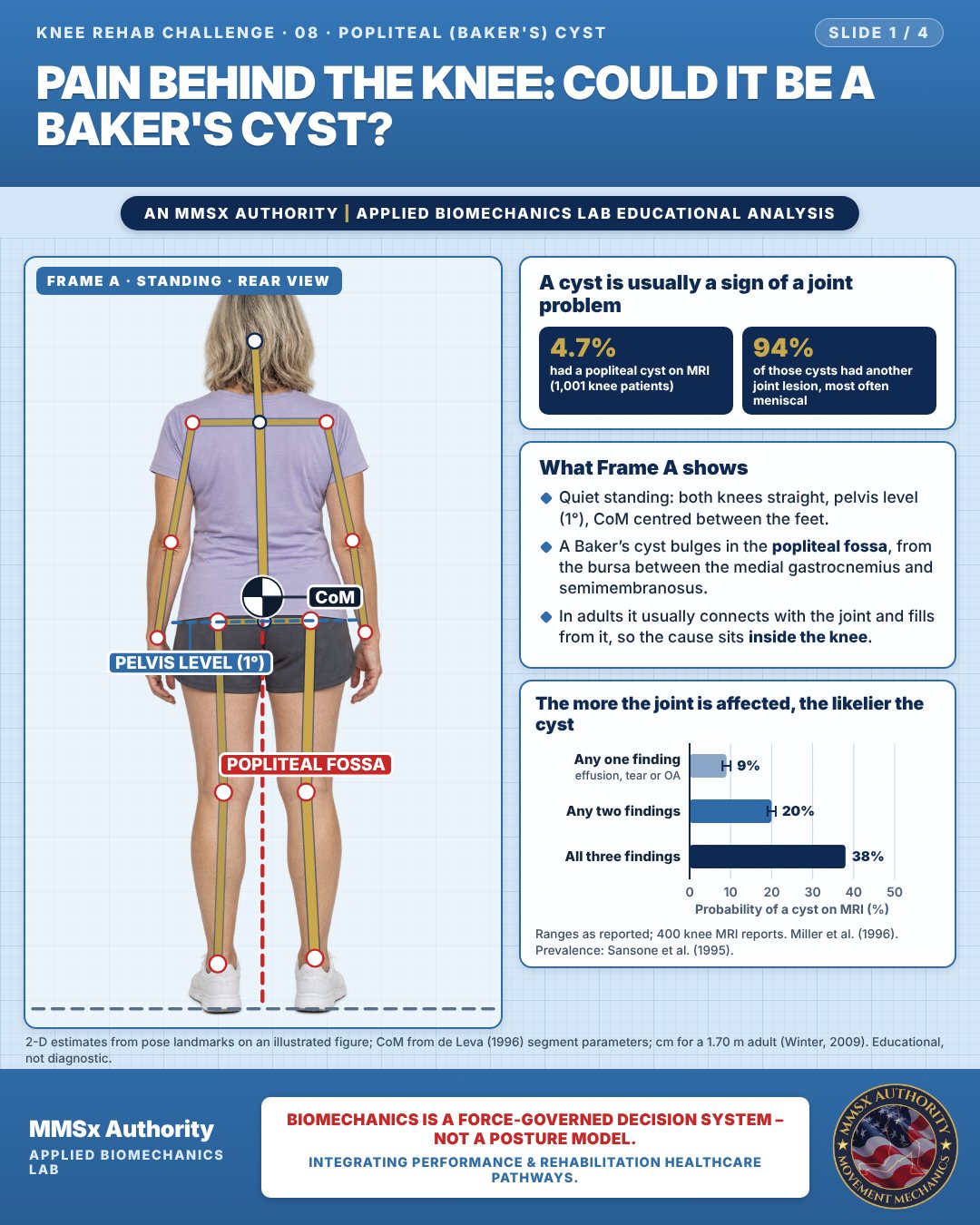
A cyst is usually a sign of a joint problem
What Frame A shows
Quiet standing: both knees straight, pelvis level (1°), CoM centred between the feet.
A Baker’s cyst bulges in the popliteal fossa, from the bursa between the medial gastrocnemius and semimembranosus.
In adults it usually connects with the joint and fills from it, so the cause sits inside the knee.
The more the joint is affected, the likelier the cyst
Movement B: Walking, late stance (side view)
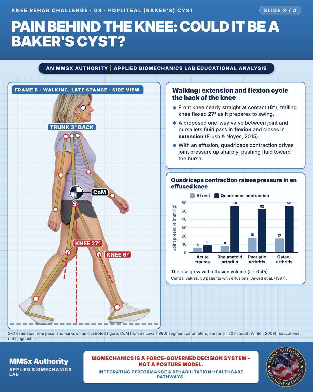
Walking: extension and flexion cycle the back of the knee
Front knee nearly straight at contact (6°); trailing knee flexed 27° as it prepares to swing.
A proposed one-way valve between joint and bursa lets fluid pass in flexion and closes in extension (Frush & Noyes, 2015).
With an effusion, quadriceps contraction drives joint pressure up sharply, pushing fluid toward the bursa.
Quadriceps contraction raises pressure in an effused knee
The rise grew with effusion volume (r = 0.45).
Movement C: Seated, knee bent, rear-oblique
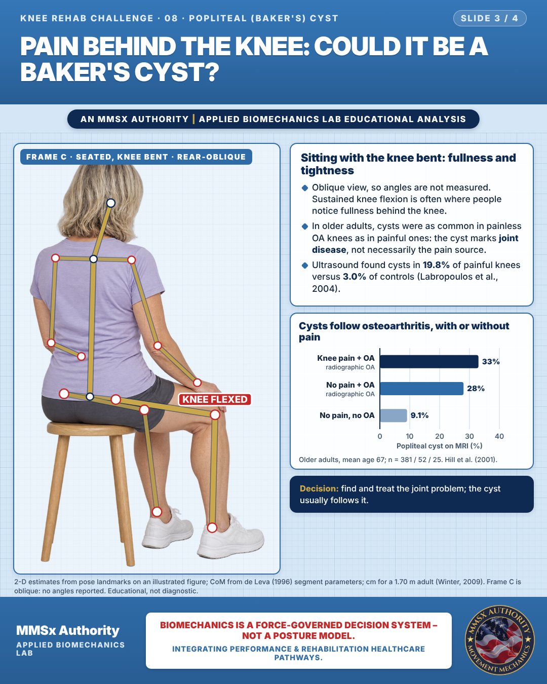
Sitting with the knee bent: fullness and tightness
Oblique view, so angles are not measured. Sustained knee flexion is often where people notice fullness behind the knee.
In older adults, cysts were as common in painless OA knees as in painful ones: the cyst marks joint disease, not necessarily the pain source.
Ultrasound found cysts in 19.8% of painful knees versus 3.0% of controls (Labropoulos et al., 2004).
Cysts follow osteoarthritis, with or without pain
Decision pathway
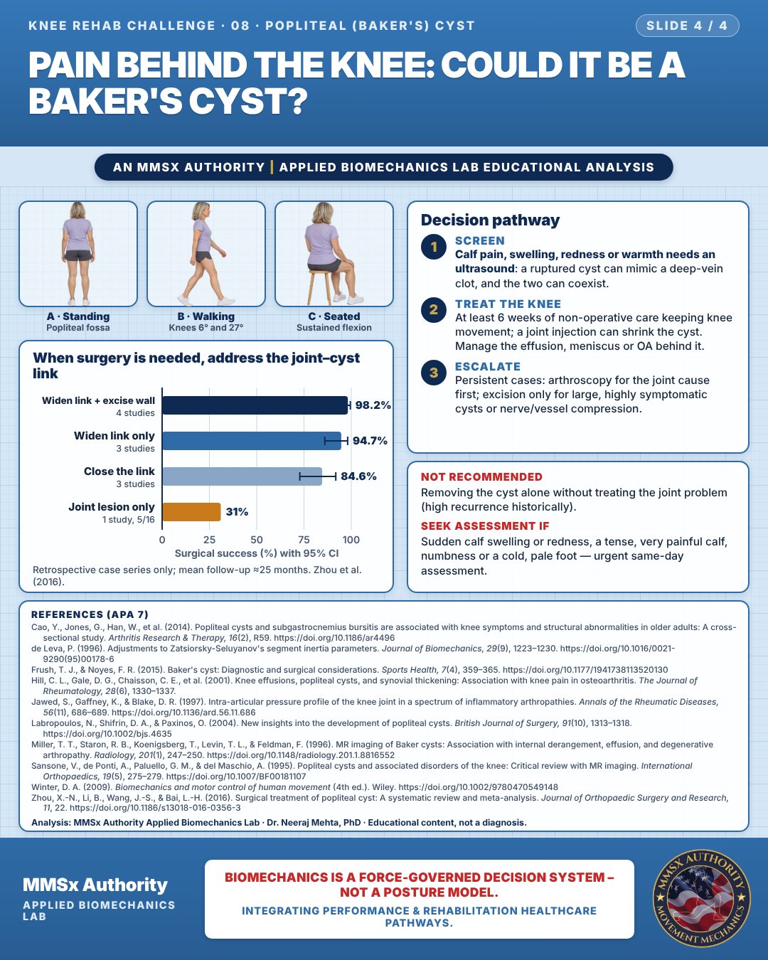
When surgery is needed, address the joint–cyst link
Screen
Calf pain, swelling, redness or warmth needs an ultrasound: a ruptured cyst can mimic a deep-vein clot, and the two can coexist.
Treat the knee
At least 6 weeks of non-operative care keeping knee movement; a joint injection can shrink the cyst. Manage the effusion, meniscus or OA behind it.
Escalate
Persistent cases: arthroscopy for the joint cause first; excision only for large, highly symptomatic cysts or nerve/vessel compression.
What the evidence supports
- Recommended: initial non-operative care for at least 6 weeks, maintaining knee flexibility; intra-articular corticosteroid can reduce cyst size and symptoms; if persistent, surgery directed at the intra-articular cause first (arthroscopy); cyst excision reserved for very large, highly symptomatic cysts or neurovascular compression (Frush & Noyes, 2015).
- Not recommended / low value: isolated cyst excision without addressing intra-articular pathology (historically high recurrence — 63% after open excision in a 1979 series of 40, reported in Zhou et al., 2016); arthroscopic communication closure has lower pooled success than enlargement (Zhou et al., 2016).
Myth and evidence
Myth“The lump is the problem — drain or cut it out.”
Evidence~94% of cysts accompany another joint lesion (Sansone et al., 1995); cyst probability rises with the number of intra-articular findings (Miller et al., 1996); outcomes are best when the joint problem and the cyst–joint valve are both addressed (Zhou et al., 2016).
Myth“Calf swelling with a known Baker's cyst is just the cyst.”
EvidenceClinical signs cannot separate rupture from DVT — scan (Frush & Noyes, 2015).
Cautions
Not recommended
Removing the cyst alone without treating the joint problem (high recurrence historically).
Seek assessment if
Sudden calf swelling or redness, a tense, very painful calf, numbness or a cold, pale foot — urgent same-day assessment.
Methods note
2-D estimates from pose landmarks on an illustrated figure; CoM from de Leva (1996) segment parameters; cm for a 1.70 m adult (Winter, 2009). Frame C is oblique: no angles reported. These are educational estimates, not a diagnosis.
References
- Cao, Y., Jones, G., Han, W., et al. (2014). Popliteal cysts and subgastrocnemius bursitis are associated with knee symptoms and structural abnormalities in older adults: A cross-sectional study. Arthritis Research & Therapy, 16(2), R59. https://doi.org/10.1186/ar4496
- de Leva, P. (1996). Adjustments to Zatsiorsky-Seluyanov's segment inertia parameters. Journal of Biomechanics, 29(9), 1223–1230. https://doi.org/10.1016/0021-9290(95)00178-6
- Frush, T. J., & Noyes, F. R. (2015). Baker's cyst: Diagnostic and surgical considerations. Sports Health, 7(4), 359–365. https://doi.org/10.1177/1941738113520130
- Hill, C. L., Gale, D. G., Chaisson, C. E., et al. (2001). Knee effusions, popliteal cysts, and synovial thickening: Association with knee pain in osteoarthritis. The Journal of Rheumatology, 28(6), 1330–1337.
- Jawed, S., Gaffney, K., & Blake, D. R. (1997). Intra-articular pressure profile of the knee joint in a spectrum of inflammatory arthropathies. Annals of the Rheumatic Diseases, 56(11), 686–689. https://doi.org/10.1136/ard.56.11.686
- Labropoulos, N., Shifrin, D. A., & Paxinos, O. (2004). New insights into the development of popliteal cysts. British Journal of Surgery, 91(10), 1313–1318. https://doi.org/10.1002/bjs.4635
- Miller, T. T., Staron, R. B., Koenigsberg, T., Levin, T. L., & Feldman, F. (1996). MR imaging of Baker cysts: Association with internal derangement, effusion, and degenerative arthropathy. Radiology, 201(1), 247–250. https://doi.org/10.1148/radiology.201.1.8816552
- Sansone, V., de Ponti, A., Paluello, G. M., & del Maschio, A. (1995). Popliteal cysts and associated disorders of the knee: Critical review with MR imaging. International Orthopaedics, 19(5), 275–279. https://doi.org/10.1007/BF00181107
- Winter, D. A. (2009). Biomechanics and motor control of human movement (4th ed.). Wiley. https://doi.org/10.1002/9780470549148
- Zhou, X.-N., Li, B., Wang, J.-S., & Bai, L.-H. (2016). Surgical treatment of popliteal cyst: A systematic review and meta-analysis. Journal of Orthopaedic Surgery and Research, 11, 22. https://doi.org/10.1186/s13018-016-0356-3
Cite this article
Mehta, N. (2026, October 23). Pain behind the knee: could it be a Baker's cyst? MMSx Authority Applied Biomechanics Lab. https://mmsxauthority.org/edu/analysis/knee/08-pain-behind-the-knee-could-it-be-a-bakers-cyst/
Part of the collection
Mehta, N. (2026). Knee Rehabilitation: 50 Evidence-Based Biomechanics Analyses [Collection]. MMSx Authority Applied Biomechanics Lab. Zenodo. https://doi.org/10.5281/zenodo.23735752