Knee Rehab Challenge · 07
Inner knee pain: why the location matters
Inner (medial) knee pain is a symptom location, not a diagnosis. Movement analysis, verified research data and a decision pathway from the MMSx Authority Appl
Key points
- Locate. Joint line pain after a twist suggests meniscus; just below the joint line on the tibia suggests pes anserine; gradual pain with stiffness suggests OA.
- Match the plan. OA: exercise and weight management. Degenerative tear: exercise first (Kise et al., 2016). Pes anserine: load change and progressive strengthening.
- Dose the task. Adjust step height, squat depth and volume; progress as symptoms allow.
What it is
Inner (medial) knee pain is a symptom location, not a diagnosis; common sources include medial tibiofemoral osteoarthritis, medial meniscal tears, medial collateral ligament (MCL) sprain and pes anserine bursitis/tendinopathy, and the precise site, onset (trauma vs gradual) and provoking tasks help separate them.
How we analysed the movement
Three movements relevant to this topic were analysed from illustrated reference figures. Joint centres were located with two-dimensional pose estimation; joint angles, the whole-body centre of mass and, where relevant, the ground-reaction-force line and its moment arm about the knee were then calculated from standard segment parameters. The figures are educational estimates that show how position changes mechanical demand; they are not measurements of a patient. Every research value quoted below comes from the published source cited beside it.
Movement A: Seated reference (front view)
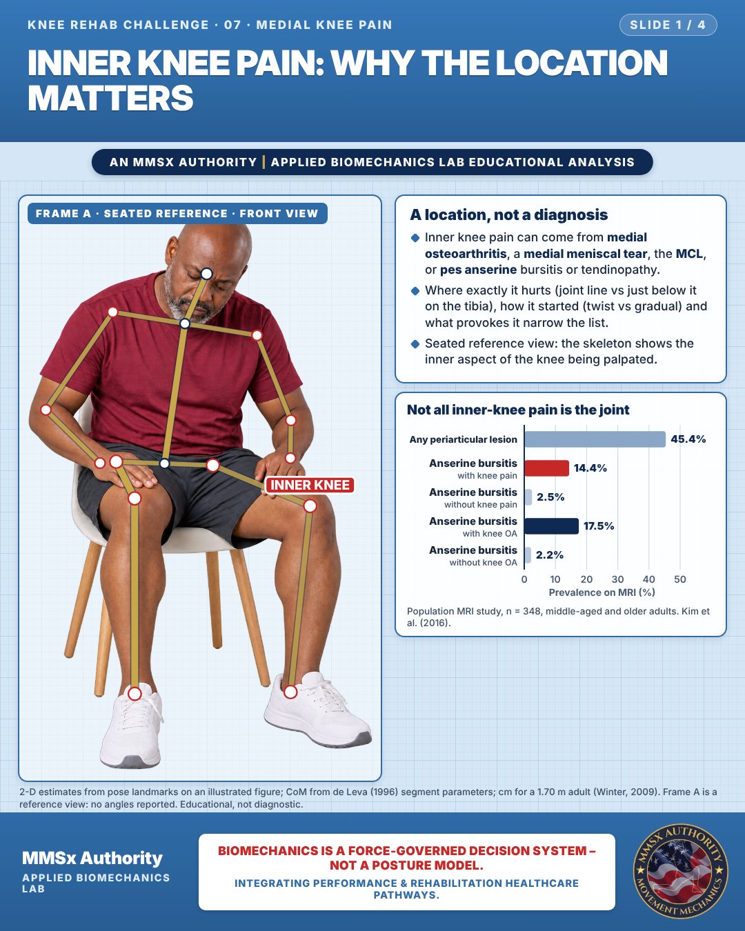
A location, not a diagnosis
Inner knee pain can come from medial osteoarthritis, a medial meniscal tear, the MCL, or pes anserine bursitis or tendinopathy.
Where exactly it hurts (joint line vs just below it on the tibia), how it started (twist vs gradual) and what provokes it narrow the list.
Seated reference view: the skeleton shows the inner aspect of the knee being palpated.
Not all inner-knee pain is the joint
Movement B: Step-up (side view)
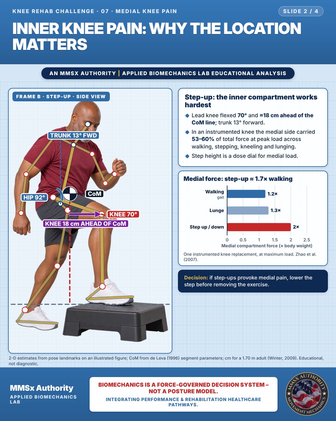
Step-up: the inner compartment works hardest
Lead knee flexed 70° and ≈18 cm ahead of the CoM line; trunk 13° forward.
In an instrumented knee the medial side carried 53–60% of total force at peak load across walking, stepping, kneeling and lunging.
Step height is a dose dial for medial load.
Medial force: step-up ≈ 1.7× walking
Movement C: Shallow squat (front view)
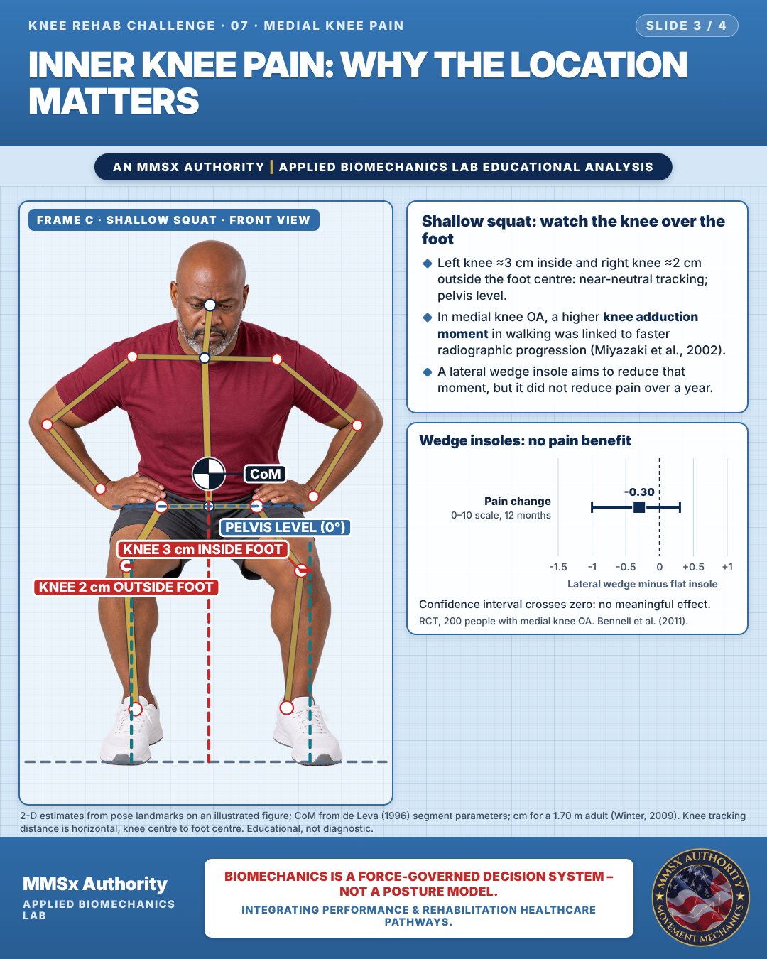
Shallow squat: watch the knee over the foot
Left knee ≈3 cm inside and right knee ≈2 cm outside the foot centre: near-neutral tracking; pelvis level.
In medial knee OA, a higher knee adduction moment in walking was linked to faster radiographic progression (Miyazaki et al., 2002).
A lateral wedge insole aims to reduce that moment, but it did not reduce pain over a year.
Wedge insoles: no pain benefit
Confidence interval crosses zero: no meaningful effect.
Decision pathway
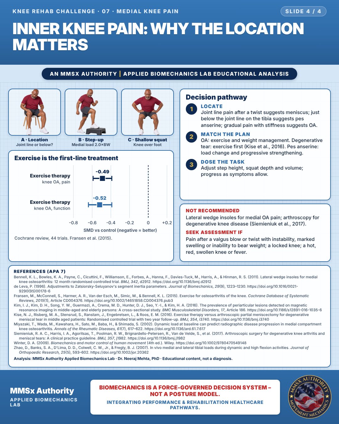
Exercise is the first-line treatment
Locate
Joint line pain after a twist suggests meniscus; just below the joint line on the tibia suggests pes anserine; gradual pain with stiffness suggests OA.
Match the plan
OA: exercise and weight management. Degenerative tear: exercise first (Kise et al., 2016). Pes anserine: load change and progressive strengthening.
Dose the task
Adjust step height, squat depth and volume; progress as symptoms allow.
Myth and evidence
Myth“A wedge insole will offload the inner knee and fix the pain.”
Evidence— no pain benefit over 12 months (−0.3/10, CI −1.0 to 0.3; Bennell et al., 2011).
Cautions
Not recommended
Lateral wedge insoles for medial OA pain; arthroscopy for degenerative knee disease (Siemieniuk et al., 2017).
Seek assessment if
Pain after a valgus blow or twist with instability, marked swelling or inability to bear weight; a locked knee; a hot, red, swollen knee or fever.
Methods note
2-D estimates from pose landmarks on an illustrated figure; CoM from de Leva (1996) segment parameters; cm for a 1.70 m adult (Winter, 2009). Frame A is a reference view: no angles reported. Knee tracking distance is horizontal, knee centre to foot centre. These are educational estimates, not a diagnosis.
References
- Bennell, K. L., Bowles, K. A., Payne, C., Cicuttini, F., Williamson, E., Forbes, A., Hanna, F., Davies-Tuck, M., Harris, A., & Hinman, R. S. (2011). Lateral wedge insoles for medial knee osteoarthritis: 12 month randomised controlled trial. BMJ, 342, d2912. https://doi.org/10.1136/bmj.d2912
- de Leva, P. (1996). Adjustments to Zatsiorsky-Seluyanov's segment inertia parameters. Journal of Biomechanics, 29(9), 1223–1230. https://doi.org/10.1016/0021-9290(95)00178-6
- Fransen, M., McConnell, S., Harmer, A. R., Van der Esch, M., Simic, M., & Bennell, K. L. (2015). Exercise for osteoarthritis of the knee. Cochrane Database of Systematic Reviews, 2015(1), Article CD004376. https://doi.org/10.1002/14651858.CD004376.pub3
- Kim, I. J., Kim, D. H., Song, Y. W., Guermazi, A., Crema, M. D., Hunter, D. J., Seo, Y.-I., & Kim, H. A. (2016). The prevalence of periarticular lesions detected on magnetic resonance imaging in middle-aged and elderly persons: A cross-sectional study. BMC Musculoskeletal Disorders, 17, Article 186. https://doi.org/10.1186/s12891-016-1035-6
- Kise, N. J., Risberg, M. A., Stensrud, S., Ranstam, J., Engebretsen, L., & Roos, E. M. (2016). Exercise therapy versus arthroscopic partial meniscectomy for degenerative meniscal tear in middle aged patients: Randomised controlled trial with two year follow-up. BMJ, 354, i3740. https://doi.org/10.1136/bmj.i3740
- Miyazaki, T., Wada, M., Kawahara, H., Sato, M., Baba, H., & Shimada, S. (2002). Dynamic load at baseline can predict radiographic disease progression in medial compartment knee osteoarthritis. Annals of the Rheumatic Diseases, 61(7), 617–622. https://doi.org/10.1136/ard.61.7.617
- Siemieniuk, R. A. C., Harris, I. A., Agoritsas, T., Poolman, R. W., Brignardello-Petersen, R., Van de Velde, S., et al. (2017). Arthroscopic surgery for degenerative knee arthritis and meniscal tears: A clinical practice guideline. BMJ, 357, j1982. https://doi.org/10.1136/bmj.j1982
- Winter, D. A. (2009). Biomechanics and motor control of human movement (4th ed.). Wiley. https://doi.org/10.1002/9780470549148
- Zhao, D., Banks, S. A., D'Lima, D. D., Colwell, C. W., Jr., & Fregly, B. J. (2007). In vivo medial and lateral tibial loads during dynamic and high flexion activities. Journal of Orthopaedic Research, 25(5), 593–602. https://doi.org/10.1002/jor.20362
Cite this article
Mehta, N. (2026, October 23). Inner knee pain: why the location matters. MMSx Authority Applied Biomechanics Lab. https://mmsxauthority.org/edu/analysis/knee/07-inner-knee-pain-why-the-location-matters/
Part of the collection
Mehta, N. (2026). Knee Rehabilitation: 50 Evidence-Based Biomechanics Analyses [Collection]. MMSx Authority Applied Biomechanics Lab. Zenodo. https://doi.org/10.5281/zenodo.23735752