Knee Rehab Challenge · 02
Knee osteoarthritis: why everyday movements become difficult
Knee osteoarthritis (OA) is a whole-joint condition involving articular cartilage loss, bone remodelling, osteophytes and synovial change.
Key points
- Diagnose. Clinically: age ≥45, activity-related pain, morning stiffness ≤30 min. Routine imaging is not needed (NICE, 2022).
- Build. Therapeutic exercise for everyone: strengthening plus aerobic fitness, supervised where possible. If overweight, any weight loss helps; 10% beats 5%.
- Keep going. Practise the hard tasks — sit-to-stand and stairs — progressing height and repetitions. Benefits fade when exercise stops.
What it is
Knee osteoarthritis (OA) is a whole-joint condition involving articular cartilage loss, bone remodelling, osteophytes and synovial change; NICE allows a clinical diagnosis without imaging in people aged 45 or over with activity-related joint pain and either no morning stiffness or morning stiffness lasting no longer than 30 minutes (NICE NG226, rec 1.1.1).
How we analysed the movement
Three movements relevant to this topic were analysed from illustrated reference figures. Joint centres were located with two-dimensional pose estimation; joint angles, the whole-body centre of mass and, where relevant, the ground-reaction-force line and its moment arm about the knee were then calculated from standard segment parameters. The figures are educational estimates that show how position changes mechanical demand; they are not measurements of a patient. Every research value quoted below comes from the published source cited beside it.
Movement A: Sit-to-stand (side view)
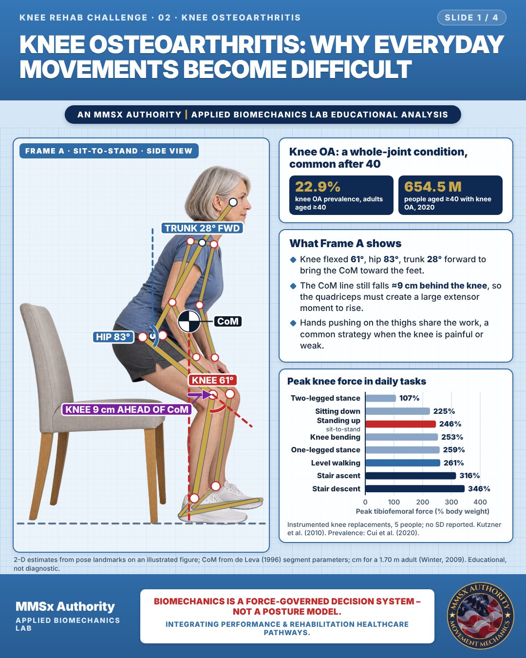
Knee OA: a whole-joint condition, common after 40
What Frame A shows
Knee flexed 61°, hip 83°, trunk 28° forward to bring the CoM toward the feet.
The CoM line still falls ≈9 cm behind the knee, so the quadriceps must create a large extensor moment to rise.
Hands pushing on the thighs share the work, a common strategy when the knee is painful or weak.
Peak knee force in daily tasks
Movement B: Stair ascent with rail (side view)
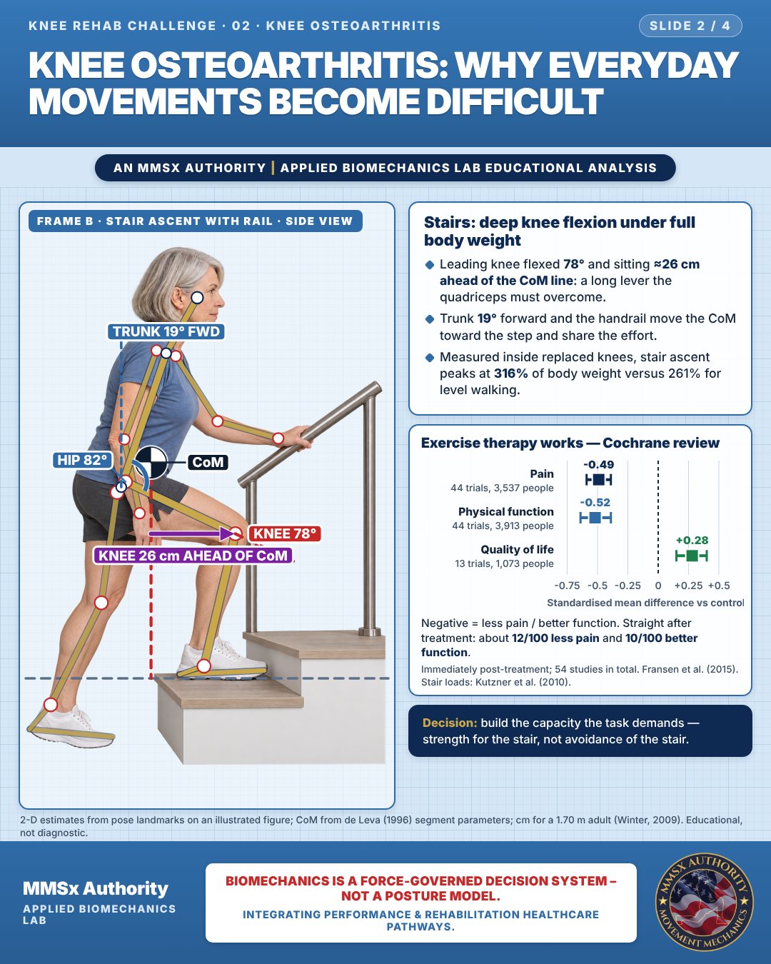
Stairs: deep knee flexion under full body weight
Leading knee flexed 78° and sitting ≈26 cm ahead of the CoM line: a long lever the quadriceps must overcome.
Trunk 19° forward and the handrail move the CoM toward the step and share the effort.
Measured inside replaced knees, stair ascent peaks at 316% of body weight versus 261% for level walking.
Exercise therapy works — Cochrane review
Negative = less pain / better function. Straight after treatment: about 12/100 less pain and 10/100 better function.
Movement C: Walking (oblique view)
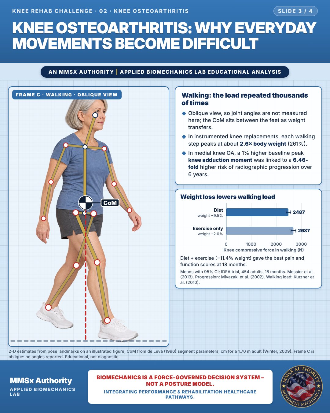
Walking: the load repeated thousands of times
Oblique view, so joint angles are not measured here; the CoM sits between the feet as weight transfers.
In instrumented knee replacements, each walking step peaks at about 2.6× body weight (261%).
In medial knee OA, a 1% higher baseline peak knee adduction moment was linked to a 6.46-fold higher risk of radiographic progression over 6 years.
Weight loss lowers walking load
Diet + exercise (−11.4% weight) gave the best pain and function scores at 18 months.
Decision pathway
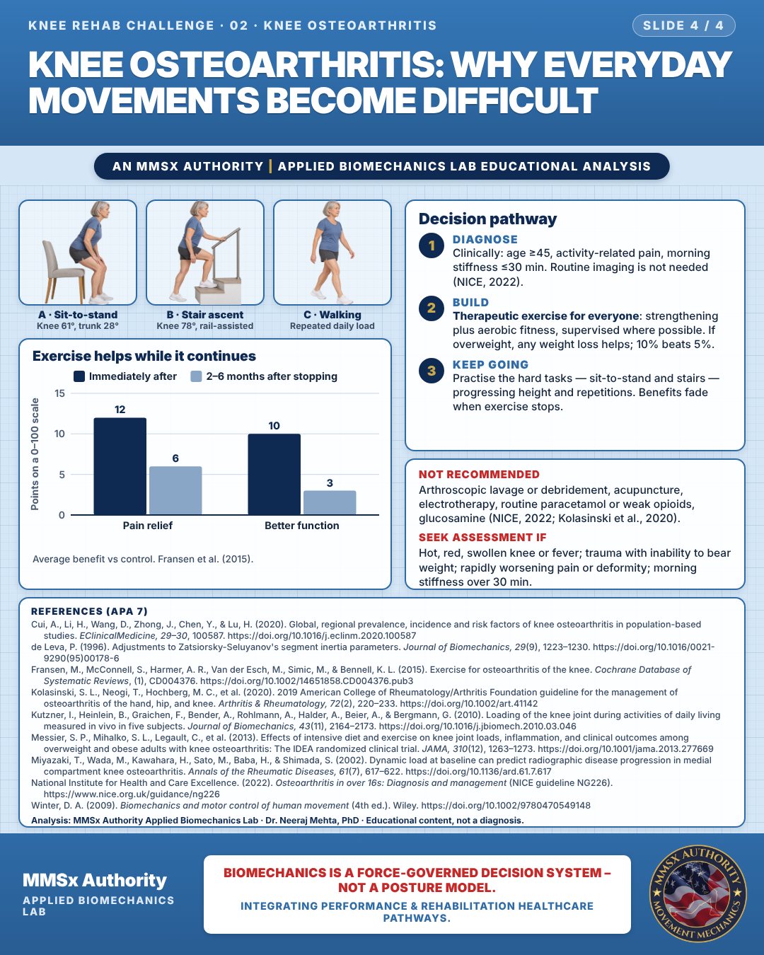
Exercise helps while it continues
Diagnose
Clinically: age ≥45, activity-related pain, morning stiffness ≤30 min. Routine imaging is not needed (NICE, 2022).
Build
Therapeutic exercise for everyone: strengthening plus aerobic fitness, supervised where possible. If overweight, any weight loss helps; 10% beats 5%.
Keep going
Practise the hard tasks — sit-to-stand and stairs — progressing height and repetitions. Benefits fade when exercise stops.
Cautions
Not recommended
Arthroscopic lavage or debridement, acupuncture, electrotherapy, routine paracetamol or weak opioids, glucosamine (NICE, 2022; Kolasinski et al., 2020).
Seek assessment if
Hot, red, swollen knee or fever; trauma with inability to bear weight; rapidly worsening pain or deformity; morning stiffness over 30 min.
Methods note
2-D estimates from pose landmarks on an illustrated figure; CoM from de Leva (1996) segment parameters; cm for a 1.70 m adult (Winter, 2009). Frame C is oblique: no angles reported. These are educational estimates, not a diagnosis.
References
- Cui, A., Li, H., Wang, D., Zhong, J., Chen, Y., & Lu, H. (2020). Global, regional prevalence, incidence and risk factors of knee osteoarthritis in population-based studies. EClinicalMedicine, 29–30, 100587. https://doi.org/10.1016/j.eclinm.2020.100587
- de Leva, P. (1996). Adjustments to Zatsiorsky-Seluyanov's segment inertia parameters. Journal of Biomechanics, 29(9), 1223–1230. https://doi.org/10.1016/0021-9290(95)00178-6
- Fransen, M., McConnell, S., Harmer, A. R., Van der Esch, M., Simic, M., & Bennell, K. L. (2015). Exercise for osteoarthritis of the knee. Cochrane Database of Systematic Reviews, (1), CD004376. https://doi.org/10.1002/14651858.CD004376.pub3
- Kolasinski, S. L., Neogi, T., Hochberg, M. C., et al. (2020). 2019 American College of Rheumatology/Arthritis Foundation guideline for the management of osteoarthritis of the hand, hip, and knee. Arthritis & Rheumatology, 72(2), 220–233. https://doi.org/10.1002/art.41142
- Kutzner, I., Heinlein, B., Graichen, F., Bender, A., Rohlmann, A., Halder, A., Beier, A., & Bergmann, G. (2010). Loading of the knee joint during activities of daily living measured in vivo in five subjects. Journal of Biomechanics, 43(11), 2164–2173. https://doi.org/10.1016/j.jbiomech.2010.03.046
- Messier, S. P., Mihalko, S. L., Legault, C., et al. (2013). Effects of intensive diet and exercise on knee joint loads, inflammation, and clinical outcomes among overweight and obese adults with knee osteoarthritis: The IDEA randomized clinical trial. JAMA, 310(12), 1263–1273. https://doi.org/10.1001/jama.2013.277669
- Miyazaki, T., Wada, M., Kawahara, H., Sato, M., Baba, H., & Shimada, S. (2002). Dynamic load at baseline can predict radiographic disease progression in medial compartment knee osteoarthritis. Annals of the Rheumatic Diseases, 61(7), 617–622. https://doi.org/10.1136/ard.61.7.617
- National Institute for Health and Care Excellence. (2022). Osteoarthritis in over 16s: Diagnosis and management (NICE guideline NG226). https://www.nice.org.uk/guidance/ng226
- Winter, D. A. (2009). Biomechanics and motor control of human movement (4th ed.). Wiley. https://doi.org/10.1002/9780470549148
Cite this article
Mehta, N. (2026, October 23). Knee osteoarthritis: why everyday movements become difficult. MMSx Authority Applied Biomechanics Lab. https://mmsxauthority.org/edu/analysis/knee/02-knee-osteoarthritis-why-everyday-movements-become-difficult/
Part of the collection
Mehta, N. (2026). Knee Rehabilitation: 50 Evidence-Based Biomechanics Analyses [Collection]. MMSx Authority Applied Biomechanics Lab. Zenodo. https://doi.org/10.5281/zenodo.23735752