Knee Rehab Challenge · 01
Runner's knee: why does the front of your knee hurt?
Patellofemoral pain (often called runner's knee) is pain felt around or behind the kneecap that is typically provoked by activities that load the…
Key points
- Settle. Lower the dose, keep moving. Use tasks lower on the ladder until symptoms settle, and explain joint load to the person.
- Build. Hip- and knee-targeted exercise is the core treatment. Taping or prefabricated foot orthoses may help short term, alongside exercise.
- Return. Climb the ladder again: stairs, then running, then jumping. Runners can add gait retraining, such as a modest step-rate increase.
What it is
Patellofemoral pain (often called runner's knee) is pain felt around or behind the kneecap that is typically provoked by activities that load the patellofemoral joint with the knee bent, such as running, squatting, stair negotiation, jumping and prolonged sitting. It is a clinical diagnosis made from the history and examination once other causes of front-of-knee pain have been considered; no single structural finding explains it.
How we analysed the movement
Three movements relevant to this topic were analysed from illustrated reference figures. Joint centres were located with two-dimensional pose estimation; joint angles, the whole-body centre of mass and, where relevant, the ground-reaction-force line and its moment arm about the knee were then calculated from standard segment parameters. The figures are educational estimates that show how position changes mechanical demand; they are not measurements of a patient. Every research value quoted below comes from the published source cited beside it.
Movement A: Running, early stance (side view)
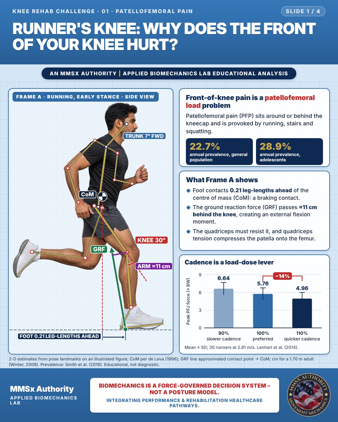
Front-of-knee pain is a patellofemoral load problem
Patellofemoral pain (PFP) sits around or behind the kneecap and is provoked by running, stairs and squatting.
What Frame A shows
Foot contacts 0.21 leg-lengths ahead of the centre of mass (CoM): a braking contact.
The ground reaction force (GRF) passes ≈11 cm behind the knee, creating an external flexion moment.
The quadriceps must resist it, and quadriceps tension compresses the patella onto the femur.
Cadence is a load-dose lever
Movement B: Step-down (side view)
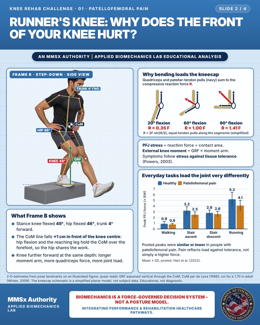
What Frame B shows
Stance knee flexed 49°, hip flexed 46°, trunk 4° forward.
The CoM line falls ≈1 cm in front of the knee centre: hip flexion and the reaching leg hold the CoM over the forefoot, so the hip shares the work.
Knee further forward at the same depth: longer moment arm, more quadriceps force, more joint load.
Why bending loads the kneecap
PFJ stress = reaction force ÷ contact area.
External knee moment = GRF × moment arm.
Symptoms follow stress against tissue tolerance (Powers, 2003).
Everyday tasks load the joint very differently
Pooled peaks were similar or lower in people with patellofemoral pain. Pain reflects load against tolerance, not simply a higher force.
Movement C: Single-leg squat (front view)
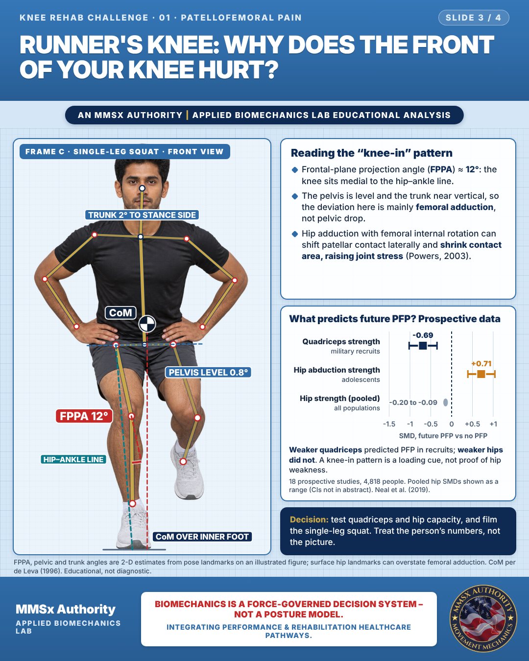
Reading the “knee-in” pattern
Frontal-plane projection angle (FPPA) ≈ 12°: the knee sits medial to the hip–ankle line.
The pelvis is level and the trunk near vertical, so the deviation here is mainly femoral adduction, not pelvic drop.
Hip adduction with femoral internal rotation can shift patellar contact laterally and shrink contact area, raising joint stress (Powers, 2003).
What predicts future PFP? Prospective data
Weaker quadriceps predicted PFP in recruits; weaker hips did not. A knee-in pattern is a loading cue, not proof of hip weakness.
Decision pathway
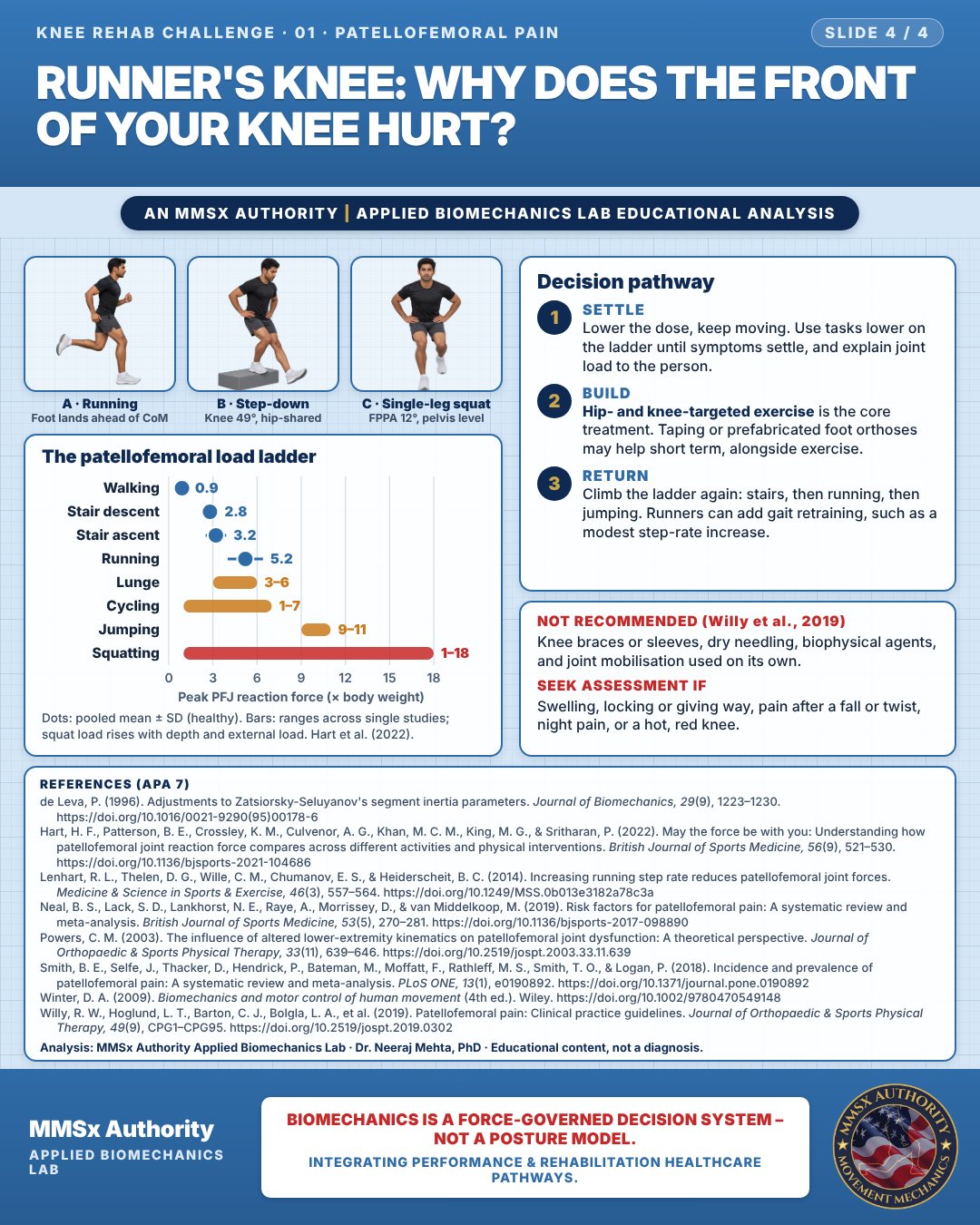
The patellofemoral load ladder
Settle
Lower the dose, keep moving. Use tasks lower on the ladder until symptoms settle, and explain joint load to the person.
Build
Hip- and knee-targeted exercise is the core treatment. Taping or prefabricated foot orthoses may help short term, alongside exercise.
Return
Climb the ladder again: stairs, then running, then jumping. Runners can add gait retraining, such as a modest step-rate increase.
Cautions
Not recommended
Knee braces or sleeves, dry needling, biophysical agents, and joint mobilisation used on its own.
Seek assessment if
Swelling, locking or giving way, pain after a fall or twist, night pain, or a hot, red knee.
Methods note
2-D estimates from pose landmarks on an illustrated figure; CoM per de Leva (1996); GRF line approximated contact point → CoM; cm for a 1.70 m adult (Winter, 2009). Prevalence: Smith et al. (2018). 2-D estimates from pose landmarks on an illustrated figure; quasi-static GRF assumed vertical through the CoM; CoM per de Leva (1996); cm for a 1.70 m adult (Winter, 2009). The kneecap schematic is a simplified planar model, not subject data. FPPA, pelvic and trunk angles are 2-D estimates from pose landmarks on an illustrated figure; surface hip landmarks can overstate femoral adduction. CoM per de Leva (1996). These are educational estimates, not a diagnosis.
References
- de Leva, P. (1996). Adjustments to Zatsiorsky-Seluyanov's segment inertia parameters. Journal of Biomechanics, 29(9), 1223–1230. https://doi.org/10.1016/0021-9290(95)00178-6
- Hart, H. F., Patterson, B. E., Crossley, K. M., Culvenor, A. G., Khan, M. C. M., King, M. G., & Sritharan, P. (2022). May the force be with you: Understanding how patellofemoral joint reaction force compares across different activities and physical interventions. British Journal of Sports Medicine, 56(9), 521–530. https://doi.org/10.1136/bjsports-2021-104686
- Lenhart, R. L., Thelen, D. G., Wille, C. M., Chumanov, E. S., & Heiderscheit, B. C. (2014). Increasing running step rate reduces patellofemoral joint forces. Medicine & Science in Sports & Exercise, 46(3), 557–564. https://doi.org/10.1249/MSS.0b013e3182a78c3a
- Neal, B. S., Lack, S. D., Lankhorst, N. E., Raye, A., Morrissey, D., & van Middelkoop, M. (2019). Risk factors for patellofemoral pain: A systematic review and meta-analysis. British Journal of Sports Medicine, 53(5), 270–281. https://doi.org/10.1136/bjsports-2017-098890
- Powers, C. M. (2003). The influence of altered lower-extremity kinematics on patellofemoral joint dysfunction: A theoretical perspective. Journal of Orthopaedic & Sports Physical Therapy, 33(11), 639–646. https://doi.org/10.2519/jospt.2003.33.11.639
- Smith, B. E., Selfe, J., Thacker, D., Hendrick, P., Bateman, M., Moffatt, F., Rathleff, M. S., Smith, T. O., & Logan, P. (2018). Incidence and prevalence of patellofemoral pain: A systematic review and meta-analysis. PLoS ONE, 13(1), e0190892. https://doi.org/10.1371/journal.pone.0190892
- Winter, D. A. (2009). Biomechanics and motor control of human movement (4th ed.). Wiley. https://doi.org/10.1002/9780470549148
- Willy, R. W., Hoglund, L. T., Barton, C. J., Bolgla, L. A., et al. (2019). Patellofemoral pain: Clinical practice guidelines. Journal of Orthopaedic & Sports Physical Therapy, 49(9), CPG1–CPG95. https://doi.org/10.2519/jospt.2019.0302
Cite this article
Mehta, N. (2026, October 23). Runner's knee: why does the front of your knee hurt? MMSx Authority Applied Biomechanics Lab. https://mmsxauthority.org/edu/analysis/knee/01-runners-knee-why-does-the-front-of-your-knee-hurt/
Part of the collection
Mehta, N. (2026). Knee Rehabilitation: 50 Evidence-Based Biomechanics Analyses [Collection]. MMSx Authority Applied Biomechanics Lab. Zenodo. https://doi.org/10.5281/zenodo.23735752