Hip & Groin Rehab Challenge · 09
Hip labral tear: when a scan finding is not the whole story
The acetabular labrum is a fibrocartilage rim around the hip socket. Movement analysis, verified research data and a decision pathway.
Key points
- Listen. Note motion- or position-related groin pain and how the person locates it, such as the C-sign.
- Examine. Use provocation tests to screen; a negative test makes joint pathology less likely, a positive one is not proof.
- Interpret. Read the scan last: treat the person, building strength and control, not the tear alone.
What it is
The acetabular labrum is a fibrocartilage rim around the hip socket. A labral tear is a structural finding on MRI, MR arthrography or at arthroscopy; it may or may not be the source of symptoms. Clinically relevant hip-related pain is typically motion- or position-related groin pain, often described with a "C-sign" around the hip, and needs to be matched with examination findings rather than diagnosed from imaging alone.
How we analysed the movement
Three movements relevant to this topic were analysed from illustrated reference figures. Joint centres were located with two-dimensional pose estimation; hip and knee angles, pelvis level, the whole-body centre of mass and, where relevant, the hip lever (the horizontal distance from the hip joint to the upper-body centre of mass) and the resulting static hip moment were then calculated from standard segment parameters. The figures are educational estimates that show how position changes mechanical demand; they are not measurements of a patient. Every research value quoted below comes from the published source cited beside it.
Movement A: Twist and pivot (side view)
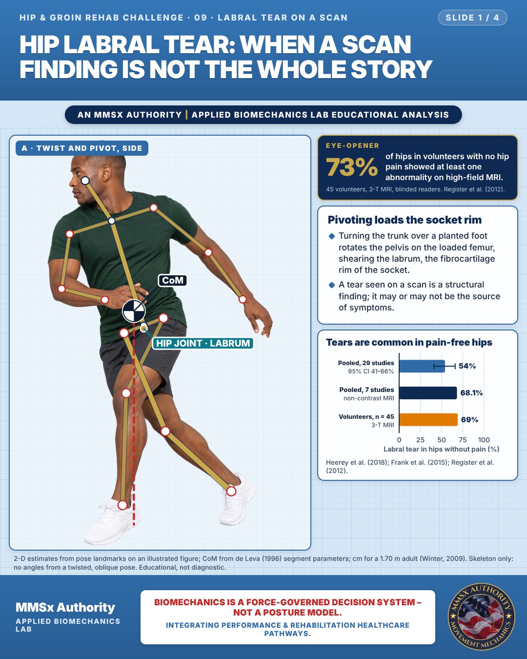
Pivoting loads the socket rim
Turning the trunk over a planted foot rotates the pelvis on the loaded femur, shearing the labrum, the fibrocartilage rim of the socket.
A tear seen on a scan is a structural finding; it may or may not be the source of symptoms.
Tears are common in pain-free hips
Movement B: Long lunge (side view)
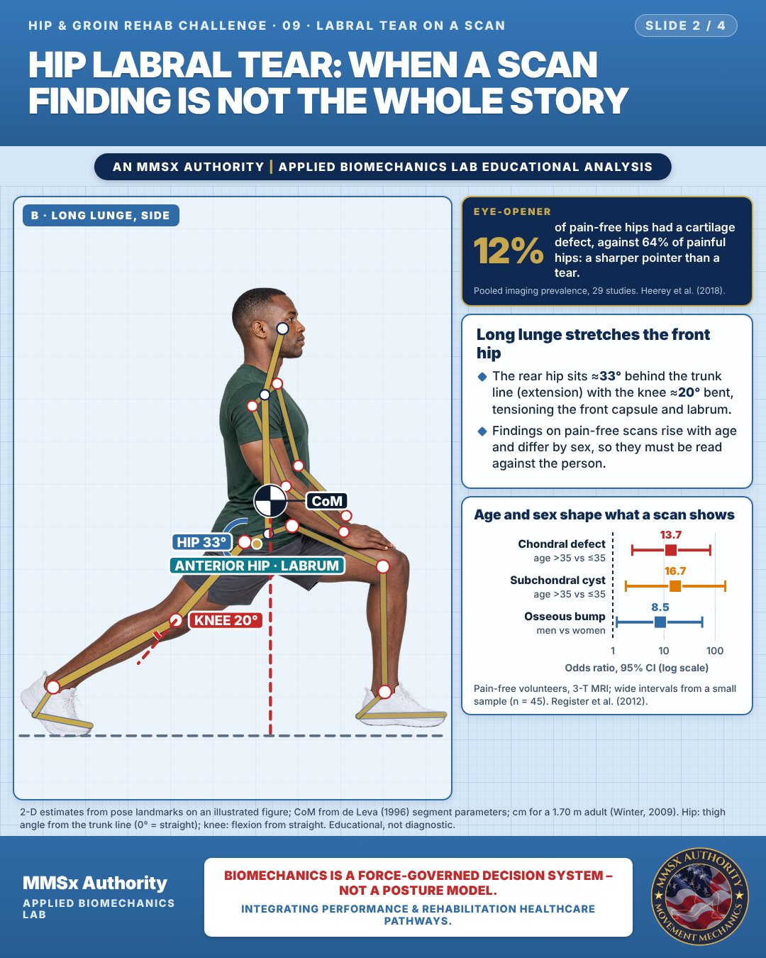
Long lunge stretches the front hip
The rear hip sits ≈33° behind the trunk line (extension) with the knee ≈20° bent, tensioning the front capsule and labrum.
Findings on pain-free scans rise with age and differ by sex, so they must be read against the person.
Age and sex shape what a scan shows
Movement C: Hand in c-shape (front view)
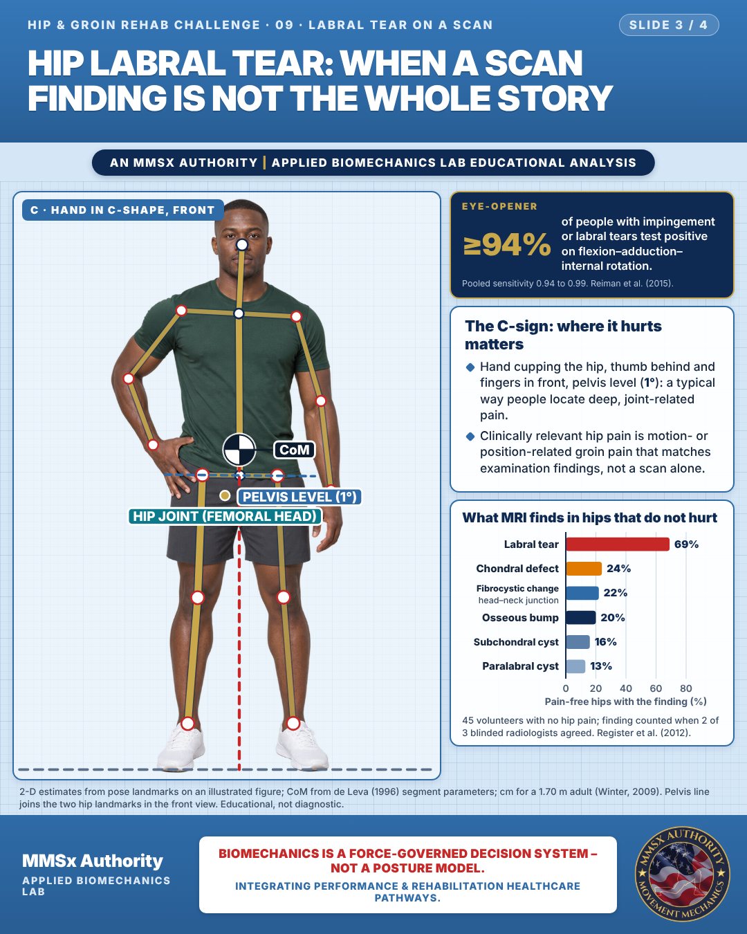
The C-sign: where it hurts matters
Hand cupping the hip, thumb behind and fingers in front, pelvis level (1°): a typical way people locate deep, joint-related pain.
Clinically relevant hip pain is motion- or position-related groin pain that matches examination findings, not a scan alone.
What MRI finds in hips that do not hurt
Decision pathway
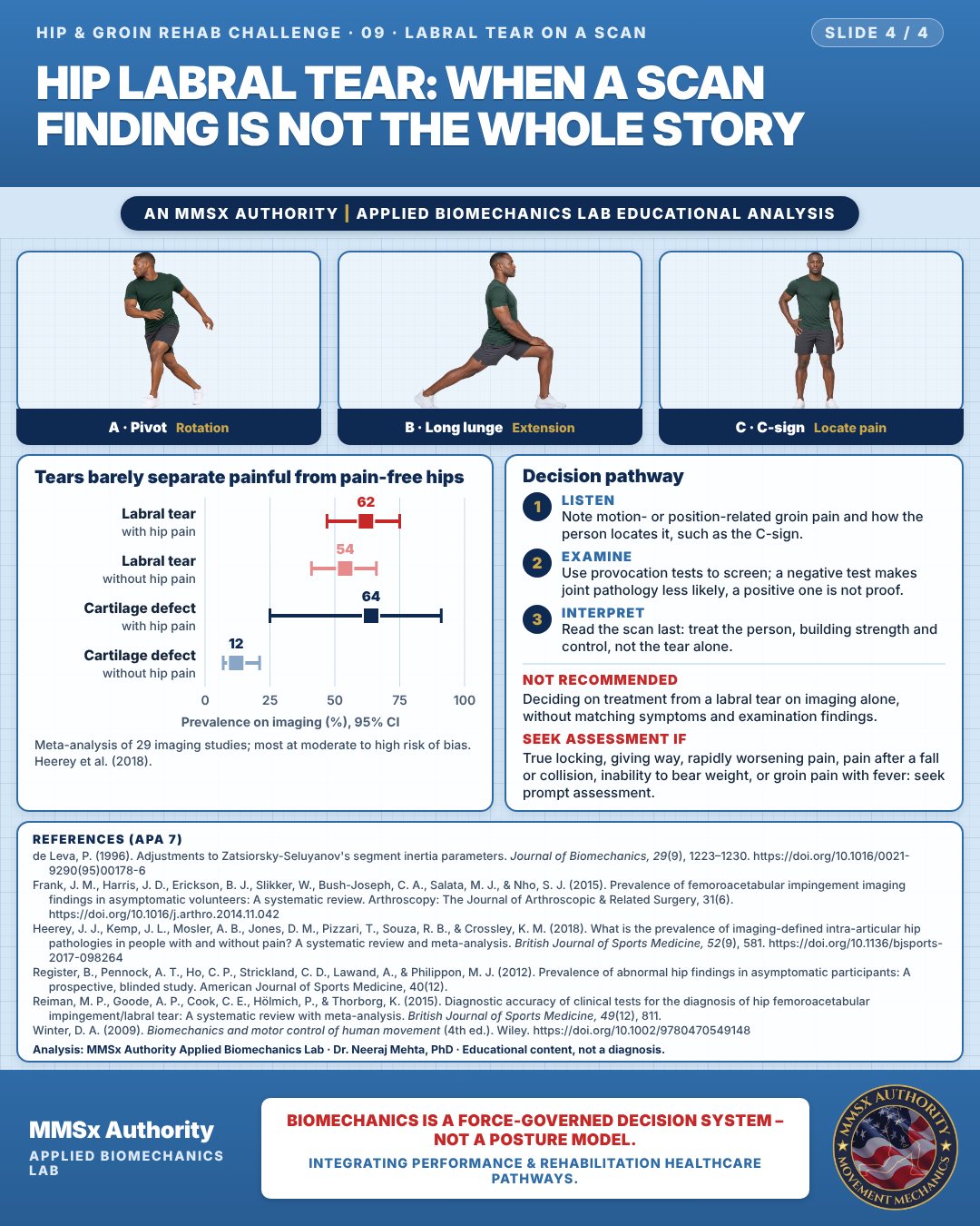
Tears barely separate painful from pain-free hips
Listen
Note motion- or position-related groin pain and how the person locates it, such as the C-sign.
Examine
Use provocation tests to screen; a negative test makes joint pathology less likely, a positive one is not proof.
Interpret
Read the scan last: treat the person, building strength and control, not the tear alone.
What the evidence supports
- Because labral tears are common in pain-free hips (54–69%), treatment decisions should not be based on imaging alone (Heerey et al., 2018; Register et al., 2012).
Myth and evidence
Myth“The MRI shows a tear, so that must be the cause of the pain.”
EvidenceLabral tears were found in 54% of people without hip pain in pooled data and in 69% of asymptomatic volunteers on 3-T MRI (Heerey et al., 2018; Register et al., 2012). Findings need to match symptoms and examination.
Cautions
Not recommended
Deciding on treatment from a labral tear on imaging alone, without matching symptoms and examination findings.
Seek assessment if
True locking, giving way, rapidly worsening pain, pain after a fall or collision, inability to bear weight, or groin pain with fever: seek prompt assessment.
Methods note
2-D estimates from pose landmarks on an illustrated figure; CoM from de Leva (1996) segment parameters; cm for a 1.70 m adult (Winter, 2009). Skeleton only: no angles from a twisted, oblique pose. Hip: thigh angle from the trunk line (0° = straight); knee: flexion from straight. Pelvis line joins the two hip landmarks in the front view. These are educational estimates, not a diagnosis.
References
- de Leva, P. (1996). Adjustments to Zatsiorsky-Seluyanov's segment inertia parameters. Journal of Biomechanics, 29(9), 1223–1230. https://doi.org/10.1016/0021-9290(95)00178-6
- Frank, J. M., Harris, J. D., Erickson, B. J., Slikker, W., Bush-Joseph, C. A., Salata, M. J., & Nho, S. J. (2015). Prevalence of femoroacetabular impingement imaging findings in asymptomatic volunteers: A systematic review. Arthroscopy: The Journal of Arthroscopic & Related Surgery, 31(6). https://doi.org/10.1016/j.arthro.2014.11.042
- Heerey, J. J., Kemp, J. L., Mosler, A. B., Jones, D. M., Pizzari, T., Souza, R. B., & Crossley, K. M. (2018). What is the prevalence of imaging-defined intra-articular hip pathologies in people with and without pain? A systematic review and meta-analysis. British Journal of Sports Medicine, 52(9), 581. https://doi.org/10.1136/bjsports-2017-098264
- Register, B., Pennock, A. T., Ho, C. P., Strickland, C. D., Lawand, A., & Philippon, M. J. (2012). Prevalence of abnormal hip findings in asymptomatic participants: A prospective, blinded study. American Journal of Sports Medicine, 40(12).
- Reiman, M. P., Goode, A. P., Cook, C. E., Hölmich, P., & Thorborg, K. (2015). Diagnostic accuracy of clinical tests for the diagnosis of hip femoroacetabular impingement/labral tear: A systematic review with meta-analysis. British Journal of Sports Medicine, 49(12), 811.
- Winter, D. A. (2009). Biomechanics and motor control of human movement (4th ed.). Wiley. https://doi.org/10.1002/9780470549148
Cite this article
Mehta, N. (2026, October 28). Hip labral tear: when a scan finding is not the whole story. MMSx Authority Applied Biomechanics Lab. https://mmsxauthority.org/edu/analysis/hip-groin/09-hip-labral-tear-when-a-scan-finding-is-not-the-whole-story/
Part of the collection
Mehta, N. (2026). Hip and Groin Rehabilitation: 30 Evidence-Based Biomechanics Analyses [Collection]. MMSx Authority Applied Biomechanics Lab. Zenodo. https://doi.org/10.5281/zenodo.23735806
All Hip & Groin Rehab Challenge articles · Rehabilitation Library