Hip & Groin Rehab Challenge · 08
Hip impingement (FAI): bone shape, movement and pain
Femoroacetabular impingement syndrome (FAI syndrome) is "a motion-related clinical disorder of the hip with a triad of symptoms, clinical signs and…
Key points
- Match. Diagnose FAI syndrome only when motion-related pain, examination signs and imaging all agree.
- Modify. Adjust depth and direction of deep flexion while rehabilitation builds strength, control and range.
- Decide. Share the choice: physiotherapist-led care or surgery, weighing a modest benefit against more adverse events.
What it is
Femoroacetabular impingement syndrome (FAI syndrome) is "a motion-related clinical disorder of the hip with a triad of symptoms, clinical signs and imaging findings". The primary symptom is motion-related or position-related pain in the hip or groin, and the imaging finding is cam and/or pincer morphology. Bone shape alone, without symptoms and signs, is not FAI syndrome (Griffin et al., 2016).
How we analysed the movement
Three movements relevant to this topic were analysed from illustrated reference figures. Joint centres were located with two-dimensional pose estimation; hip and knee angles, pelvis level, the whole-body centre of mass and, where relevant, the hip lever (the horizontal distance from the hip joint to the upper-body centre of mass) and the resulting static hip moment were then calculated from standard segment parameters. The figures are educational estimates that show how position changes mechanical demand; they are not measurements of a patient. Every research value quoted below comes from the published source cited beside it.
Movement A: Deep squat (side view)
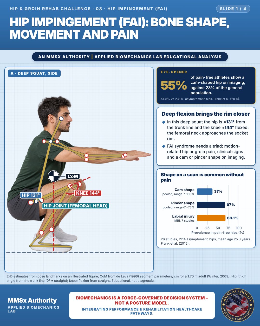
Deep flexion brings the rim closer
In this deep squat the hip is ≈131° from the trunk line and the knee ≈144° flexed: the femoral neck approaches the socket rim.
FAI syndrome needs a triad: motion-related hip or groin pain, clinical signs and a cam or pincer shape on imaging.
Shape on a scan is common without pain
Movement B: Knee across body (front view)
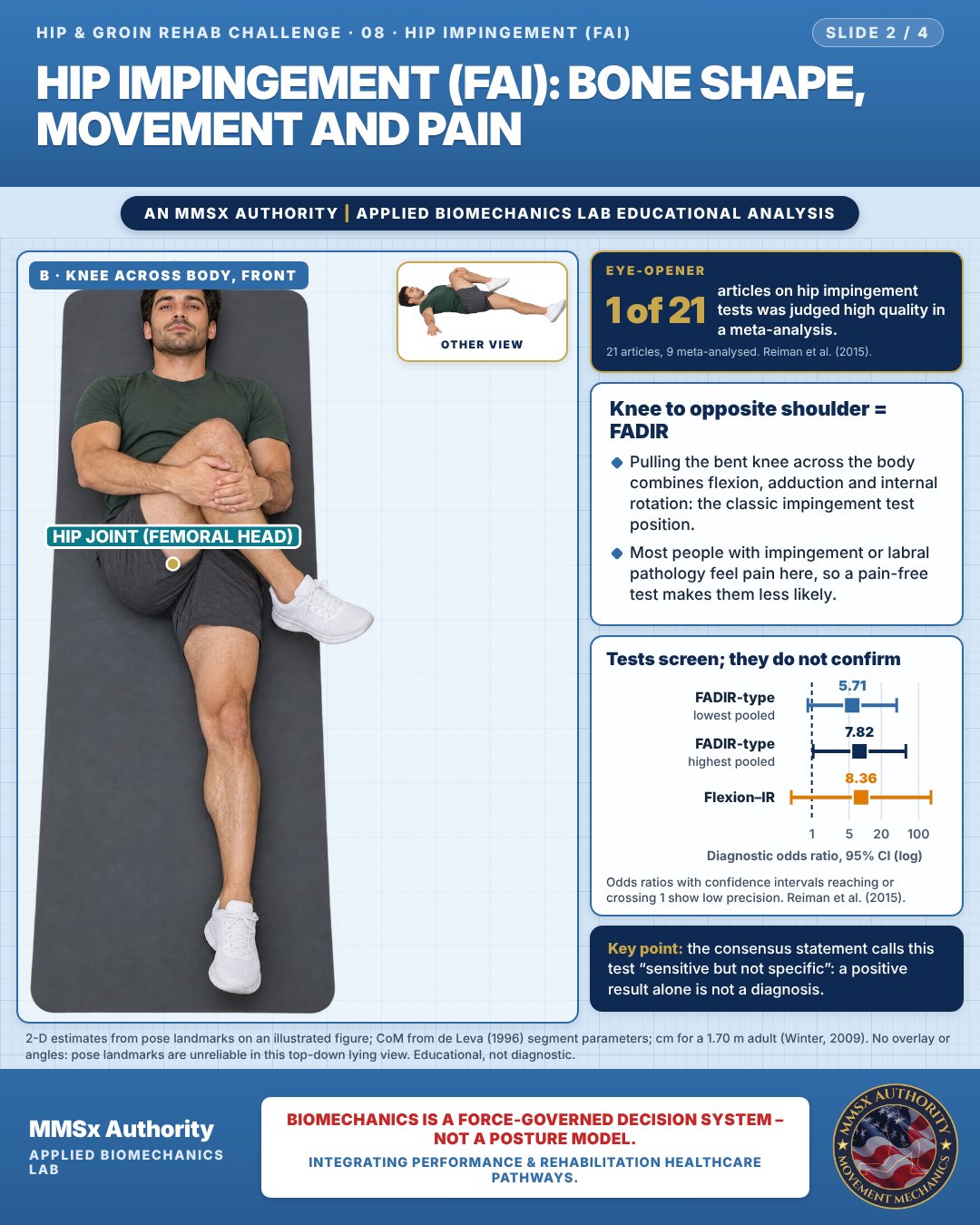
Knee to opposite shoulder = FADIR
Pulling the bent knee across the body combines flexion, adduction and internal rotation: the classic impingement test position.
Most people with impingement or labral pathology feel pain here, so a pain-free test makes them less likely.
Tests screen; they do not confirm
Movement C: High knee lift (side view)
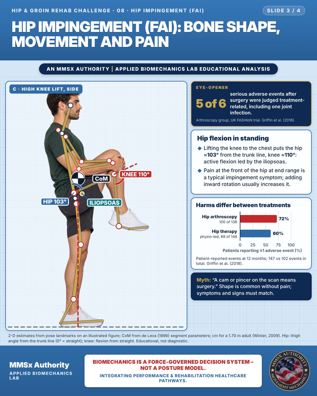
Hip flexion in standing
Lifting the knee to the chest puts the hip ≈103° from the trunk line, knee ≈110°: active flexion led by the iliopsoas.
Pain at the front of the hip at end range is a typical impingement symptom; adding inward rotation usually increases it.
Harms differ between treatments
Decision pathway
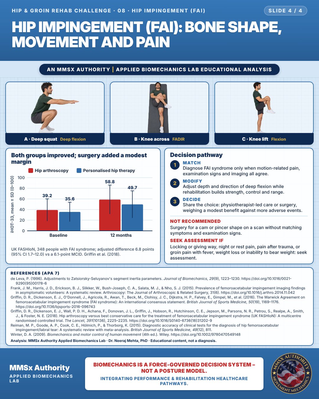
Both groups improved; surgery added a modest margin
Match
Diagnose FAI syndrome only when motion-related pain, examination signs and imaging all agree.
Modify
Adjust depth and direction of deep flexion while rehabilitation builds strength, control and range.
Decide
Share the choice: physiotherapist-led care or surgery, weighing a modest benefit against more adverse events.
What the evidence supports
- FAI syndrome "can be treated by conservative care, rehabilitation or surgery"; physiotherapy-led rehabilitation aims to improve hip stability, neuromuscular control, strength, range of motion and movement patterns; surgery (open or arthroscopic) aims to improve hip morphology and repair damaged tissue (Griffin et al., 2016).
Myth and evidence
Myth“A cam or pincer on the scan means you need surgery.”
EvidenceCam morphology is found in 37% and pincer morphology in 67% of asymptomatic hips (Frank et al., 2015), FAI syndrome needs symptoms, signs and imaging together (Griffin et al., 2016), and physiotherapist-led care helped many patients in a high-quality trial (Griffin et al., 2018).
Cautions
Not recommended
Surgery for a cam or pincer shape on a scan without matching symptoms and examination signs.
Seek assessment if
Locking or giving way, night or rest pain, pain after trauma, or groin pain with fever, weight loss or inability to bear weight: seek assessment.
Methods note
2-D estimates from pose landmarks on an illustrated figure; CoM from de Leva (1996) segment parameters; cm for a 1.70 m adult (Winter, 2009). Hip: thigh angle from the trunk line (0° = straight); knee: flexion from straight. No overlay or angles: pose landmarks are unreliable in this top-down lying view. These are educational estimates, not a diagnosis.
References
- de Leva, P. (1996). Adjustments to Zatsiorsky-Seluyanov's segment inertia parameters. Journal of Biomechanics, 29(9), 1223–1230. https://doi.org/10.1016/0021-9290(95)00178-6
- Frank, J. M., Harris, J. D., Erickson, B. J., Slikker, W., Bush-Joseph, C. A., Salata, M. J., & Nho, S. J. (2015). Prevalence of femoroacetabular impingement imaging findings in asymptomatic volunteers: A systematic review. Arthroscopy: The Journal of Arthroscopic & Related Surgery, 31(6). https://doi.org/10.1016/j.arthro.2014.11.042
- Griffin, D. R., Dickenson, E. J., O'Donnell, J., Agricola, R., Awan, T., Beck, M., Clohisy, J. C., Dijkstra, H. P., Falvey, E., Gimpel, M., et al. (2016). The Warwick Agreement on femoroacetabular impingement syndrome (FAI syndrome): An international consensus statement. British Journal of Sports Medicine, 50(19), 1169–1176. https://doi.org/10.1136/bjsports-2016-096743
- Griffin, D. R., Dickenson, E. J., Wall, P. D. H., Achana, F., Donovan, J. L., Griffin, J., Hobson, R., Hutchinson, C. E., Jepson, M., Parsons, N. R., Petrou, S., Realpe, A., Smith, J., & Foster, N. E. (2018). Hip arthroscopy versus best conservative care for the treatment of femoroacetabular impingement syndrome (UK FASHIoN): A multicentre randomised controlled trial. The Lancet, 391(10136), 2225–2235. https://doi.org/10.1016/S0140-6736(18)31202-9
- Reiman, M. P., Goode, A. P., Cook, C. E., Hölmich, P., & Thorborg, K. (2015). Diagnostic accuracy of clinical tests for the diagnosis of hip femoroacetabular impingement/labral tear: A systematic review with meta-analysis. British Journal of Sports Medicine, 49(12), 811.
- Winter, D. A. (2009). Biomechanics and motor control of human movement (4th ed.). Wiley. https://doi.org/10.1002/9780470549148
Cite this article
Mehta, N. (2026, October 28). Hip impingement (FAI): bone shape, movement and pain. MMSx Authority Applied Biomechanics Lab. https://mmsxauthority.org/edu/analysis/hip-groin/08-hip-impingement-fai-bone-shape-movement-and-pain/
Part of the collection
Mehta, N. (2026). Hip and Groin Rehabilitation: 30 Evidence-Based Biomechanics Analyses [Collection]. MMSx Authority Applied Biomechanics Lab. Zenodo. https://doi.org/10.5281/zenodo.23735806
All Hip & Groin Rehab Challenge articles · Rehabilitation Library