Hamstring & Thigh Rehab Challenge · 24
Myositis ossificans: when a thigh bruise turns to bone
Myositis ossificans (traumatic) is the formation of benign, non-cancerous bone within a muscle after injury, most often after a moderate or severe…
Key points
- Act early. Treat a heavy bruise promptly; delay over 3 days, effusion and prior injury were risk factors.
- Regain bend. Restore knee flexion gradually: seated bends, wall sits, then cycling, guided by pain.
- Image if stuck. If a firm lump persists or range stalls, imaging can confirm a benign process.
What it is
Myositis ossificans (traumatic) is the formation of benign, non-cancerous bone within a muscle after injury, most often after a moderate or severe quadriceps contusion. It presents as a painful, firm mass with persisting loss of knee bend. On plain radiographs it may be seen as early as 2 to 4 weeks after injury (Lamplot & Matava, 2016).
How we analysed the movement
Three movements relevant to this topic were analysed from illustrated reference figures. Joint centres were located with two-dimensional pose estimation; hip and knee angles, pelvis level, the whole-body centre of mass and, where relevant, the hip lever (the horizontal distance from the hip joint to the upper-body centre of mass) and the resulting static hip moment were then calculated from standard segment parameters. The figures are educational estimates that show how position changes mechanical demand; they are not measurements of a patient. Every research value quoted below comes from the published source cited beside it.
Movement A: Seated knee bend (side view)
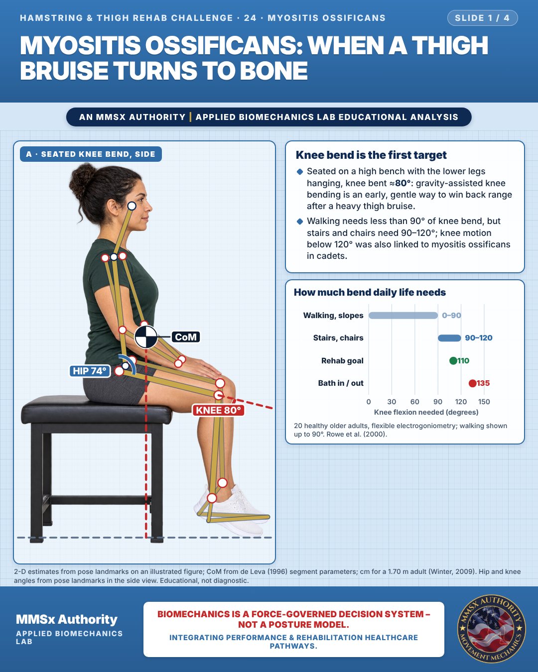
Knee bend is the first target
Seated on a high bench with the lower legs hanging, knee bent ≈80°: gravity-assisted knee bending is an early, gentle way to win back range after a heavy thigh bruise.
Walking needs less than 90° of knee bend, but stairs and chairs need 90–120°; knee motion below 120° was also linked to myositis ossificans in cadets.
How much bend daily life needs
Movement B: Wall sit (side view)
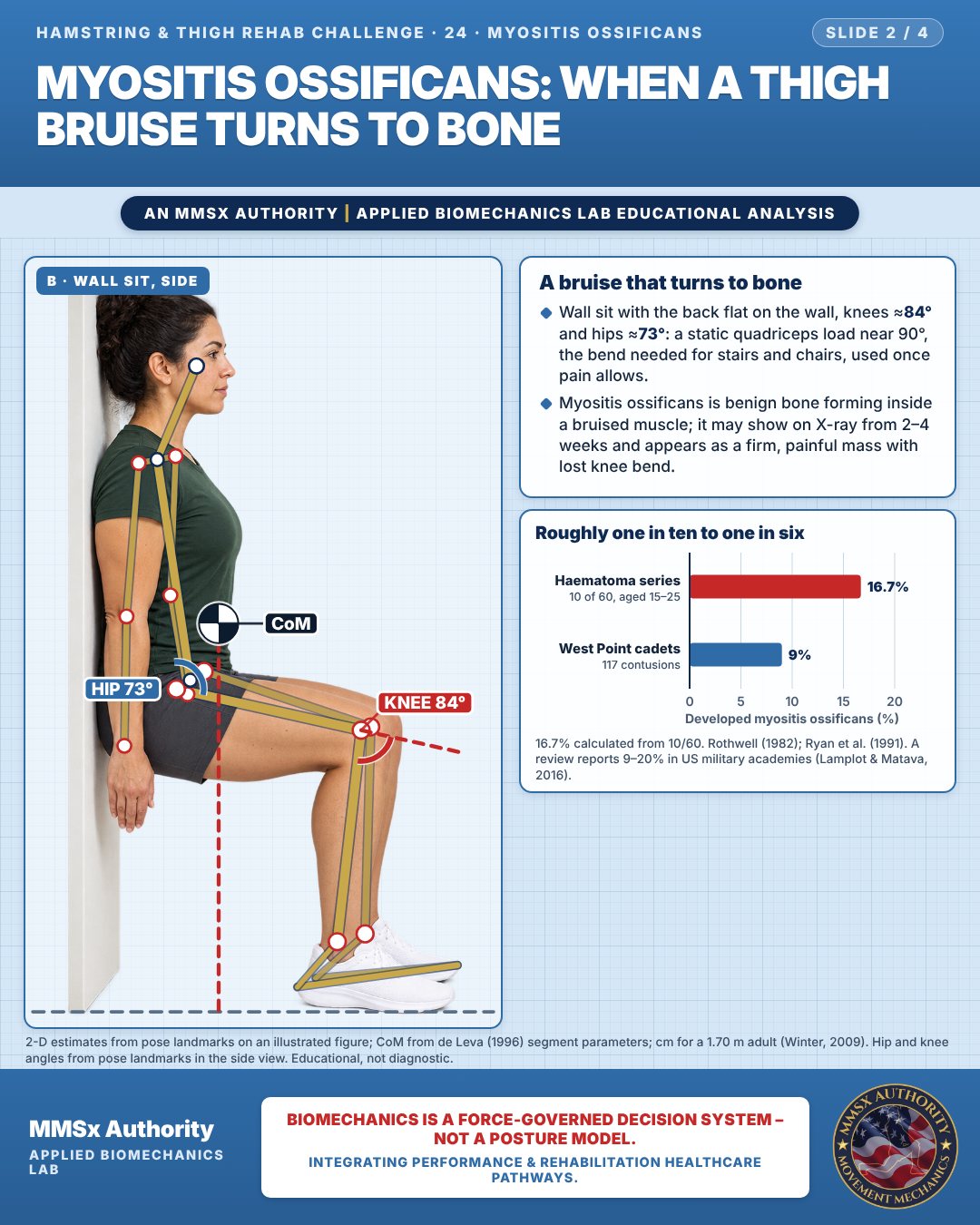
A bruise that turns to bone
Wall sit with the back flat on the wall, knees ≈84° and hips ≈73°: a static quadriceps load near 90°, the bend needed for stairs and chairs, used once pain allows.
Myositis ossificans is benign bone forming inside a bruised muscle; it may show on X-ray from 2–4 weeks and appears as a firm, painful mass with lost knee bend.
Roughly one in ten to one in six
Movement C: Stationary bike (side view)
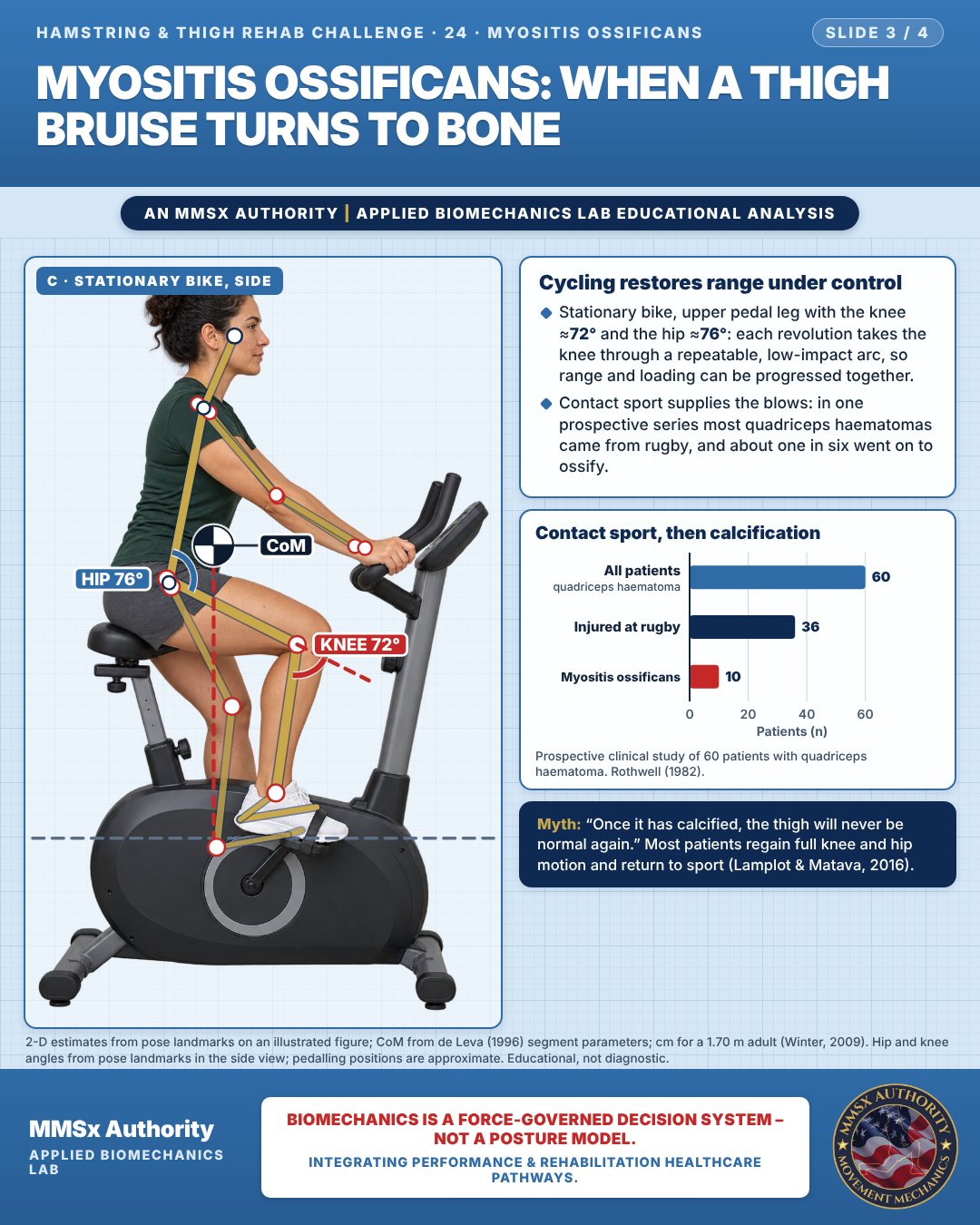
Cycling restores range under control
Stationary bike, upper pedal leg with the knee ≈72° and the hip ≈76°: each revolution takes the knee through a repeatable, low-impact arc, so range and loading can be progressed together.
Contact sport supplies the blows: in one prospective series most quadriceps haematomas came from rugby, and about one in six went on to ossify.
Contact sport, then calcification
Decision pathway
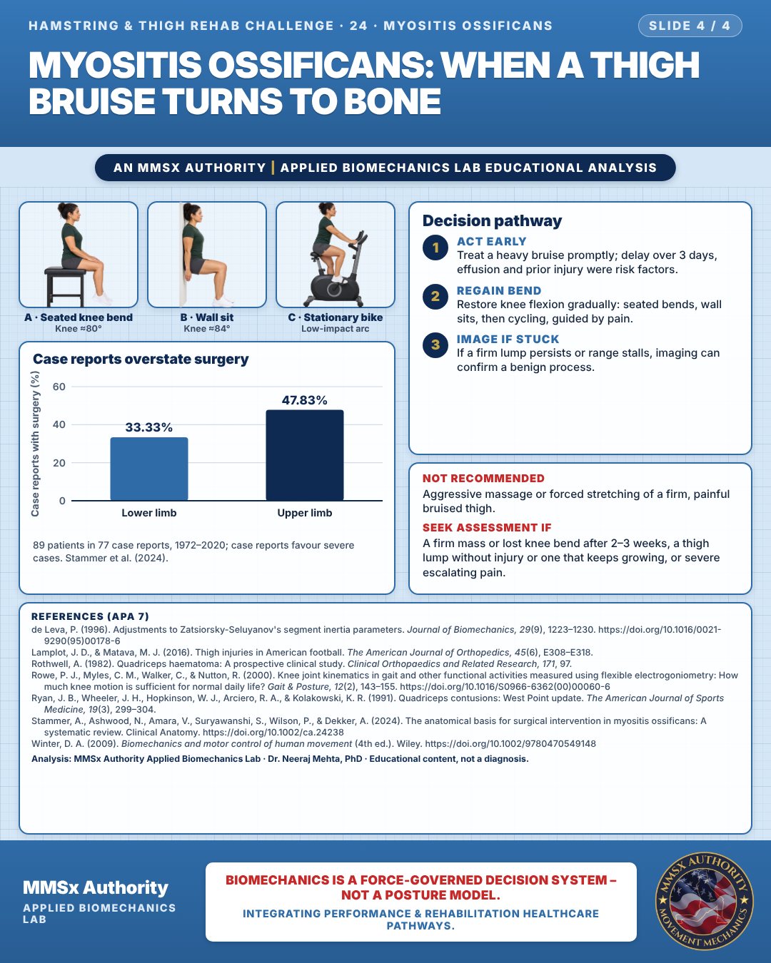
Case reports overstate surgery
Act early
Treat a heavy bruise promptly; delay over 3 days, effusion and prior injury were risk factors.
Regain bend
Restore knee flexion gradually: seated bends, wall sits, then cycling, guided by pain.
Image if stuck
If a firm lump persists or range stalls, imaging can confirm a benign process.
What the evidence supports
- Even with myositis ossificans, most patients regain full knee and hip motion and return to sport. Very rarely, persistent symptoms and limited motion warrant consideration of surgical excision (Lamplot & Matava, 2016).
- There are no data on the efficacy of NSAIDs in preventing myositis ossificans after quadriceps contusions (Lamplot & Matava, 2016).
- Practical implication: early, pain-guided restoration of knee flexion and gradual loading (seated knee bends, wall sits, then stationary cycling) is consistent with the three-phase approach (Ryan et al., 1991).
Myth and evidence
Myth“Once the bruise has calcified, the thigh will never be normal again.”
EvidenceMost patients with myositis ossificans regain full knee and hip motion and return to sport, and surgery is very rarely needed (Lamplot & Matava, 2016).
Cautions
Not recommended
Aggressive massage or forced stretching of a firm, painful bruised thigh.
Seek assessment if
A firm mass or lost knee bend after 2–3 weeks, a thigh lump without injury or one that keeps growing, or severe escalating pain.
Methods note
2-D estimates from pose landmarks on an illustrated figure; CoM from de Leva (1996) segment parameters; cm for a 1.70 m adult (Winter, 2009). Hip and knee angles from pose landmarks in the side view. Hip and knee angles from pose landmarks in the side view; pedalling positions are approximate. These are educational estimates, not a diagnosis.
References
- de Leva, P. (1996). Adjustments to Zatsiorsky-Seluyanov's segment inertia parameters. Journal of Biomechanics, 29(9), 1223–1230. https://doi.org/10.1016/0021-9290(95)00178-6
- Lamplot, J. D., & Matava, M. J. (2016). Thigh injuries in American football. The American Journal of Orthopedics, 45(6), E308–E318.
- Rothwell, A. (1982). Quadriceps haematoma: A prospective clinical study. Clinical Orthopaedics and Related Research, 171, 97.
- Rowe, P. J., Myles, C. M., Walker, C., & Nutton, R. (2000). Knee joint kinematics in gait and other functional activities measured using flexible electrogoniometry: How much knee motion is sufficient for normal daily life? Gait & Posture, 12(2), 143–155. https://doi.org/10.1016/S0966-6362(00)00060-6
- Ryan, J. B., Wheeler, J. H., Hopkinson, W. J., Arciero, R. A., & Kolakowski, K. R. (1991). Quadriceps contusions: West Point update. The American Journal of Sports Medicine, 19(3), 299–304.
- Stammer, A., Ashwood, N., Amara, V., Suryawanshi, S., Wilson, P., & Dekker, A. (2024). The anatomical basis for surgical intervention in myositis ossificans: A systematic review. Clinical Anatomy. https://doi.org/10.1002/ca.24238
- Winter, D. A. (2009). Biomechanics and motor control of human movement (4th ed.). Wiley. https://doi.org/10.1002/9780470549148
Cite this article
Mehta, N. (2026, October 26). Myositis ossificans: when a thigh bruise turns to bone. MMSx Authority Applied Biomechanics Lab. https://mmsxauthority.org/edu/analysis/hamstring-thigh/24-myositis-ossificans-when-a-thigh-bruise-turns-to-bone/
Part of the collection
Mehta, N. (2026). Hamstring and Thigh Rehabilitation: 50 Evidence-Based Biomechanics Analyses [Collection]. MMSx Authority Applied Biomechanics Lab. Zenodo. https://doi.org/10.5281/zenodo.23735792
All Hamstring & Thigh Rehab Challenge articles · Rehabilitation Library