Hamstring & Thigh Rehab Challenge · 22
Rectus femoris injury: why the two-joint quadriceps is vulnerable
The rectus femoris is the only quadriceps muscle that crosses both the hip and the knee. Movement analysis, verified research data and a decision pathway.
Key points
- Locate. Establish the site: muscle, myotendinous junction, free tendon or bony avulsion.
- Short first. Load with the hip flexed and knee straight before combining hip extension with knee bend.
- Long last. Add half-kneeling and push-off positions, then sprinting, and kicking last.
What it is
The rectus femoris is the only quadriceps muscle that crosses both the hip and the knee. It flexes the hip and extends the knee. Injury can involve the muscle belly, the musculotendinous junction, the free proximal tendon, or the bony attachment at the anterior inferior iliac spine (avulsion). Because it spans two joints, it is lengthened most when the hip is extended and the knee flexed at the same time, as in the kicking backswing, sprint toe-off and the kneeling hip flexor stretch.
How we analysed the movement
Three movements relevant to this topic were analysed from illustrated reference figures. Joint centres were located with two-dimensional pose estimation; hip and knee angles, pelvis level, the whole-body centre of mass and, where relevant, the hip lever (the horizontal distance from the hip joint to the upper-body centre of mass) and the resulting static hip moment were then calculated from standard segment parameters. The figures are educational estimates that show how position changes mechanical demand; they are not measurements of a patient. Every research value quoted below comes from the published source cited beside it.
Movement A: Standing quad stretch (side view)
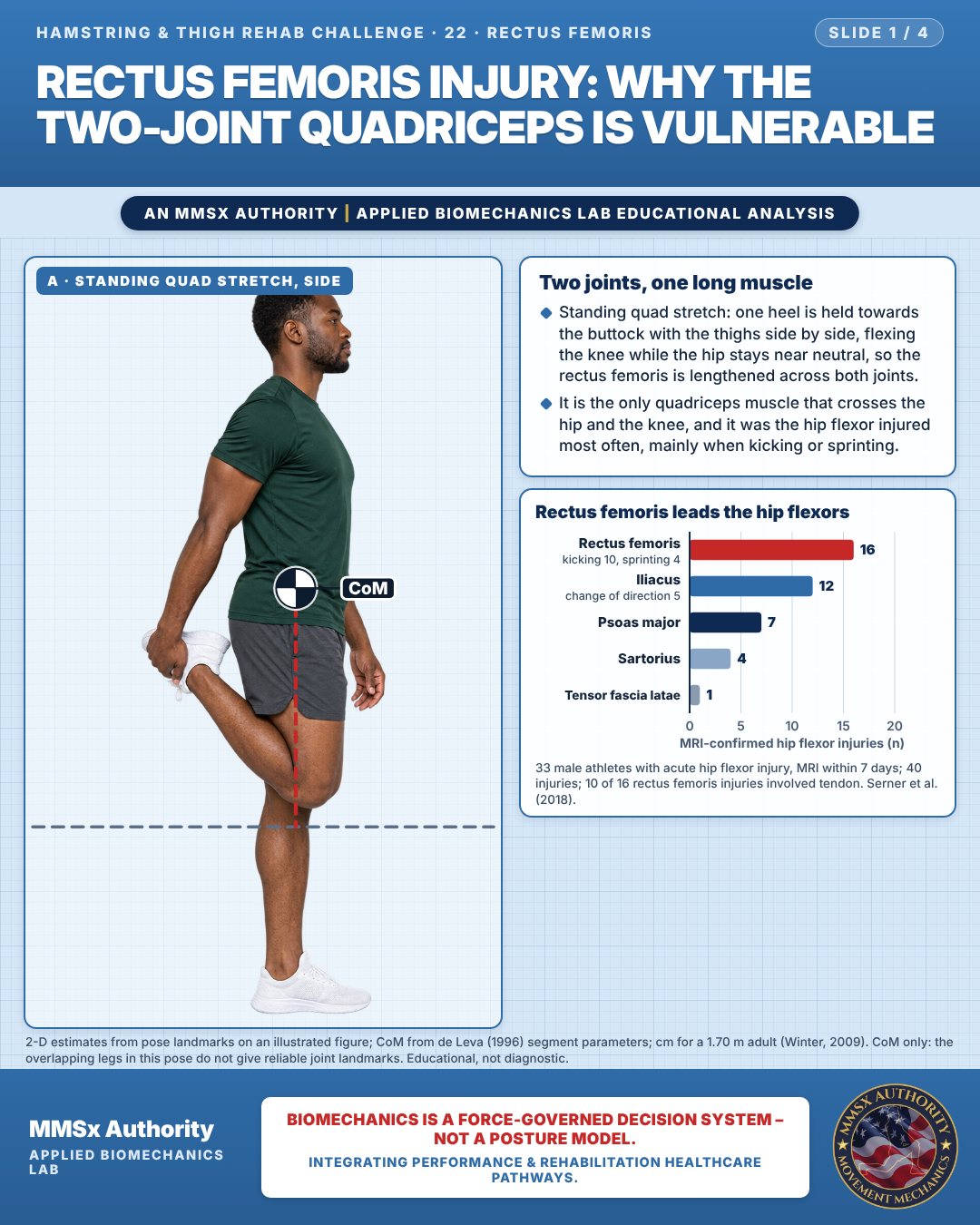
Two joints, one long muscle
Standing quad stretch: one heel is held towards the buttock with the thighs side by side, flexing the knee while the hip stays near neutral, so the rectus femoris is lengthened across both joints.
It is the only quadriceps muscle that crosses the hip and the knee, and it was the hip flexor injured most often, mainly when kicking or sprinting.
Rectus femoris leads the hip flexors
Movement B: Sprint push-off (side view)
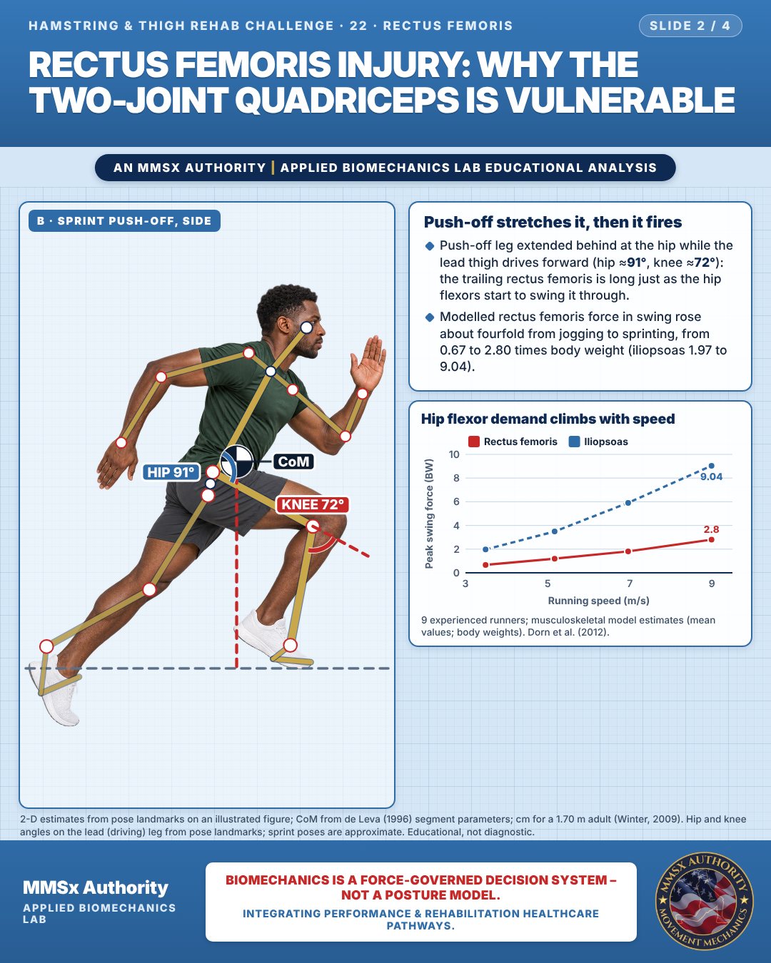
Push-off stretches it, then it fires
Push-off leg extended behind at the hip while the lead thigh drives forward (hip ≈91°, knee ≈72°): the trailing rectus femoris is long just as the hip flexors start to swing it through.
Modelled rectus femoris force in swing rose about fourfold from jogging to sprinting, from 0.67 to 2.80 times body weight (iliopsoas 1.97 to 9.04).
Hip flexor demand climbs with speed
Movement C: Half-kneeling (side view)
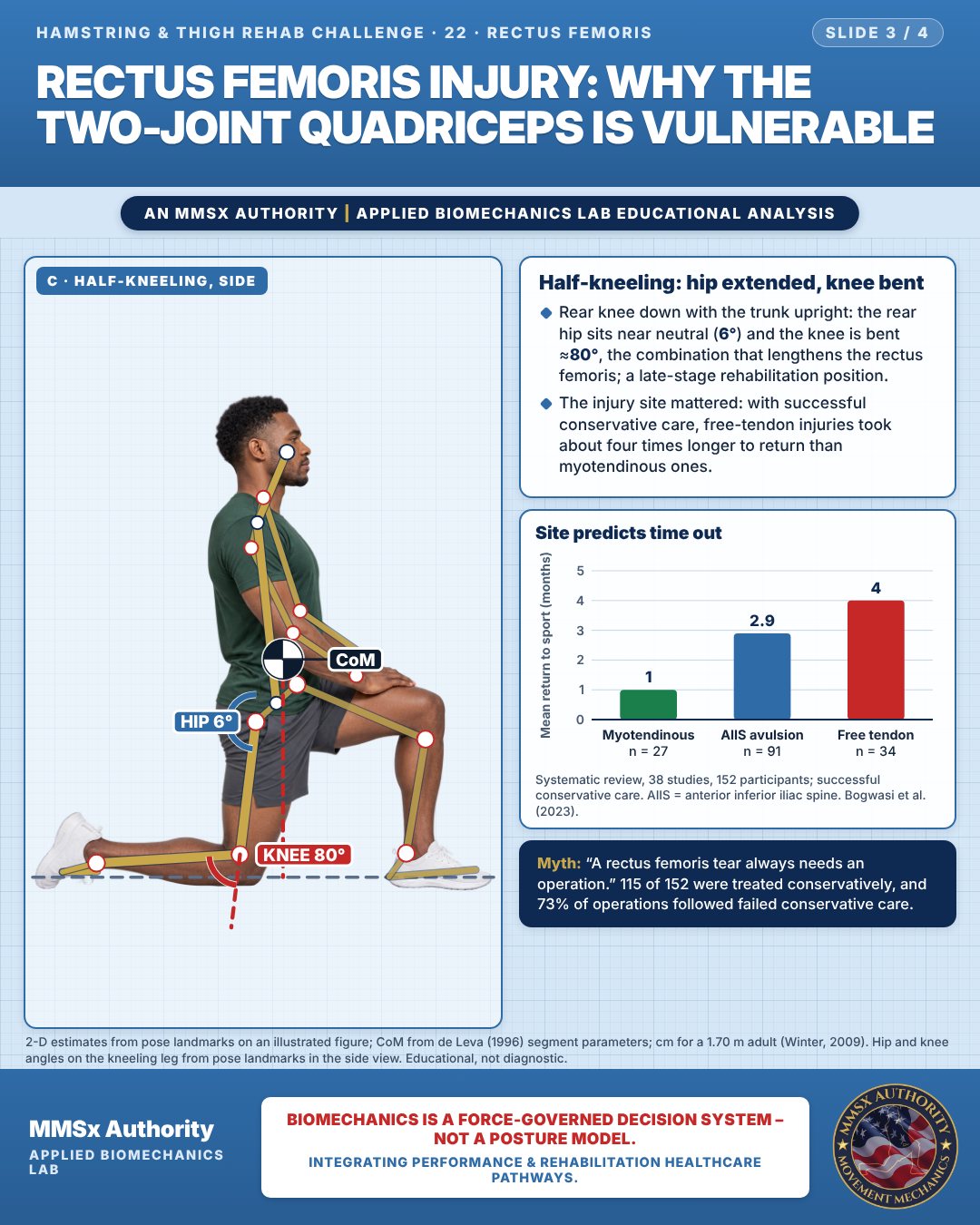
Half-kneeling: hip extended, knee bent
Rear knee down with the trunk upright: the rear hip sits near neutral (6°) and the knee is bent ≈80°, the combination that lengthens the rectus femoris; a late-stage rehabilitation position.
The injury site mattered: with successful conservative care, free-tendon injuries took about four times longer to return than myotendinous ones.
Site predicts time out
Decision pathway
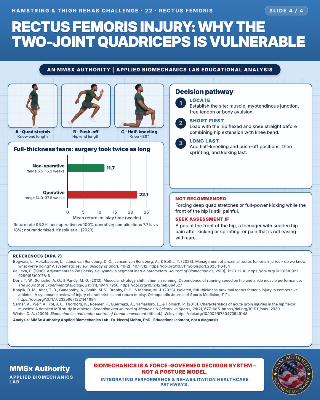
Full-thickness tears: surgery took twice as long
Locate
Establish the site: muscle, myotendinous junction, free tendon or bony avulsion.
Short first
Load with the hip flexed and knee straight before combining hip extension with knee bend.
Long last
Add half-kneeling and push-off positions, then sprinting, and kicking last.
What the evidence supports
- Most proximal rectus femoris injuries were managed conservatively. In the systematic review, 73% of surgical treatments followed failed conservative treatment, and conservative care gave the shorter mean return to sport (Bogwasi et al., 2023).
Myth and evidence
Myth“A rectus femoris tear always needs an operation.”
EvidenceMost injuries in the systematic review were treated conservatively (115 of 152), conservative care gave shorter return-to-sport times, and most surgery happened only after conservative treatment had failed (73%) (Bogwasi et al., 2023).
Cautions
Not recommended
Forcing deep quad stretches or full-power kicking while the front of the hip is still painful.
Seek assessment if
A pop at the front of the hip, a teenager with sudden hip pain after kicking or sprinting, or pain that is not easing with care.
Methods note
2-D estimates from pose landmarks on an illustrated figure; CoM from de Leva (1996) segment parameters; cm for a 1.70 m adult (Winter, 2009). CoM only: the overlapping legs in this pose do not give reliable joint landmarks. Hip and knee angles on the lead (driving) leg from pose landmarks; sprint poses are approximate. Hip and knee angles on the kneeling leg from pose landmarks in the side view. These are educational estimates, not a diagnosis.
References
- Bogwasi, L., Holtzhausen, L., Janse van Rensburg, D. C., Jansen van Rensburg, A., & Botha, T. (2023). Management of proximal rectus femoris injuries – do we know what we're doing? A systematic review. Biology of Sport, 40(2), 497–512. https://doi.org/10.5114/biolsport.2023.116454
- de Leva, P. (1996). Adjustments to Zatsiorsky-Seluyanov's segment inertia parameters. Journal of Biomechanics, 29(9), 1223–1230. https://doi.org/10.1016/0021-9290(95)00178-6
- Dorn, T. W., Schache, A. G., & Pandy, M. G. (2012). Muscular strategy shift in human running: Dependence of running speed on hip and ankle muscle performance. The Journal of Experimental Biology, 215(11), 1944–1956. https://doi.org/10.1242/jeb.064527
- Knapik, D. M., Alter, T. D., Ganapathy, A., Smith, M. V., Brophy, R. H., & Matava, M. J. (2023). Isolated, full-thickness proximal rectus femoris injury in competitive athletes: A systematic review of injury characteristics and return to play. Orthopaedic Journal of Sports Medicine, 11(1). https://doi.org/10.1177/23259671221144984
- Serner, A., Weir, A., Tol, J. L., Thorborg, K., Roemer, F., Guermazi, A., Yamashiro, E., & Hölmich, P. (2018). Characteristics of acute groin injuries in the hip flexor muscles: A detailed MRI study in athletes. Scandinavian Journal of Medicine & Science in Sports, 28(2), 677–685. https://doi.org/10.1111/sms.12939
- Winter, D. A. (2009). Biomechanics and motor control of human movement (4th ed.). Wiley. https://doi.org/10.1002/9780470549148
Cite this article
Mehta, N. (2026, October 26). Rectus femoris injury: why the two-joint quadriceps is vulnerable. MMSx Authority Applied Biomechanics Lab. https://mmsxauthority.org/edu/analysis/hamstring-thigh/22-rectus-femoris-injury-why-the-two-joint-quadriceps-is-vulnerable/
Part of the collection
Mehta, N. (2026). Hamstring and Thigh Rehabilitation: 50 Evidence-Based Biomechanics Analyses [Collection]. MMSx Authority Applied Biomechanics Lab. Zenodo. https://doi.org/10.5281/zenodo.23735792
All Hamstring & Thigh Rehab Challenge articles · Rehabilitation Library