Arm Pain Rehab Challenge · 32
Wrist sprain or scaphoid fracture? Why a “sprain” after a fall needs checking
Fracture of the scaphoid (the carpal bone on the thumb side of the wrist), typically after a fall onto the outstretched, extended hand (Movement A).
Key points
- Protect. After a fall with thumb-side pain, splint the wrist and arrange review even if the X-ray is normal.
- Check. Re-examine the snuffbox and scaphoid tubercle; positive predictors call for further imaging.
- Image. MRI combines high specificity with good sensitivity; bone scans miss little but over-treat.
What it is
Fracture of the scaphoid (the carpal bone on the thumb side of the wrist), typically after a fall onto the outstretched, extended hand (Movement A). Pain in the anatomical snuffbox (Movement B) is the key clinical sign; initial X-rays can be normal, so suspected fractures are treated and re-assessed or imaged further.
How we analysed the movement
Three movements relevant to this topic were analysed from illustrated reference figures. Joint centres were located with two-dimensional pose estimation where the whole arm was visible; elbow and wrist angles, and the directions of external load and tendon pull, were then drawn on the figure; on hand close-ups, where pose estimation does not apply, the landmarks were placed manually. The figures are educational estimates that show how position changes mechanical demand; they are not measurements of a patient. Every research value quoted below comes from the published source cited beside it.
Movement A: Fall on hands (side view)
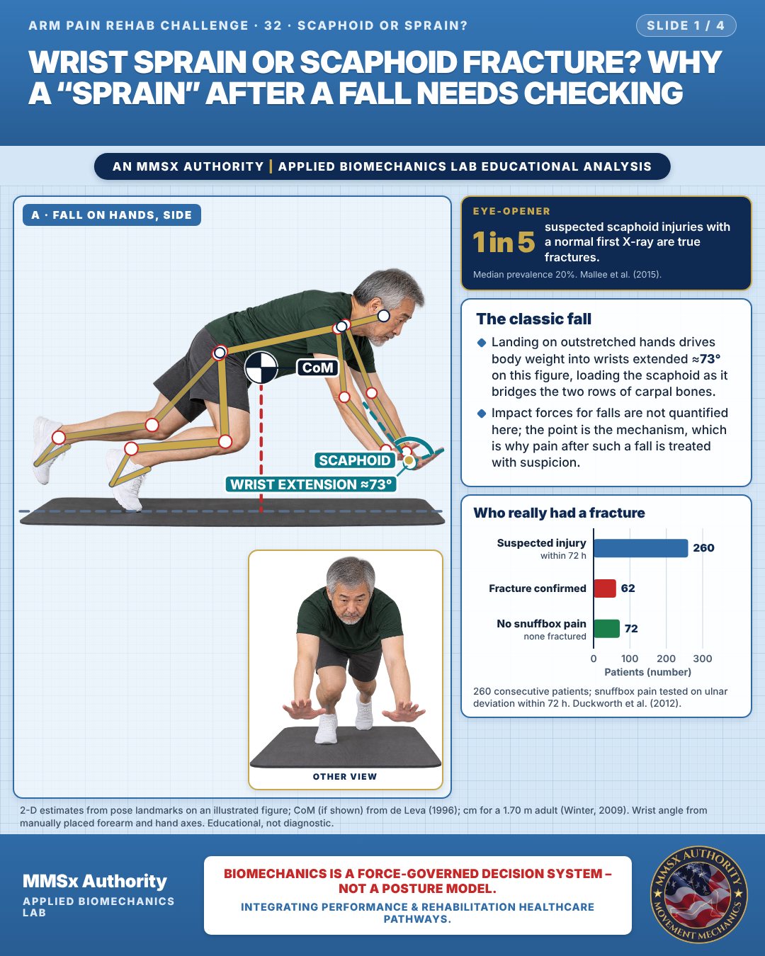
The classic fall
Landing on outstretched hands drives body weight into wrists extended ≈73° on this figure, loading the scaphoid as it bridges the two rows of carpal bones.
Impact forces for falls are not quantified here; the point is the mechanism, which is why pain after such a fall is treated with suspicion.
Who really had a fracture
Movement B: Thumb up (side view)
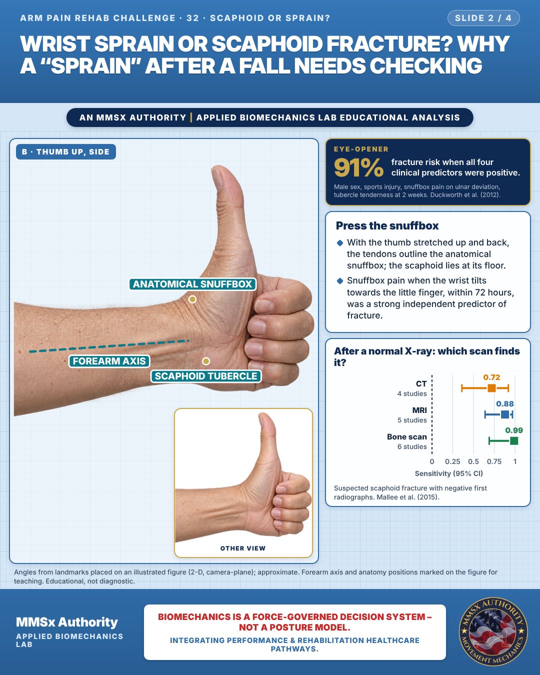
Press the snuffbox
With the thumb stretched up and back, the tendons outline the anatomical snuffbox; the scaphoid lies at its floor.
Snuffbox pain when the wrist tilts towards the little finger, within 72 hours, was a strong independent predictor of fracture.
After a normal X-ray: which scan finds it?
Movement C: Guarding wrist (front view)
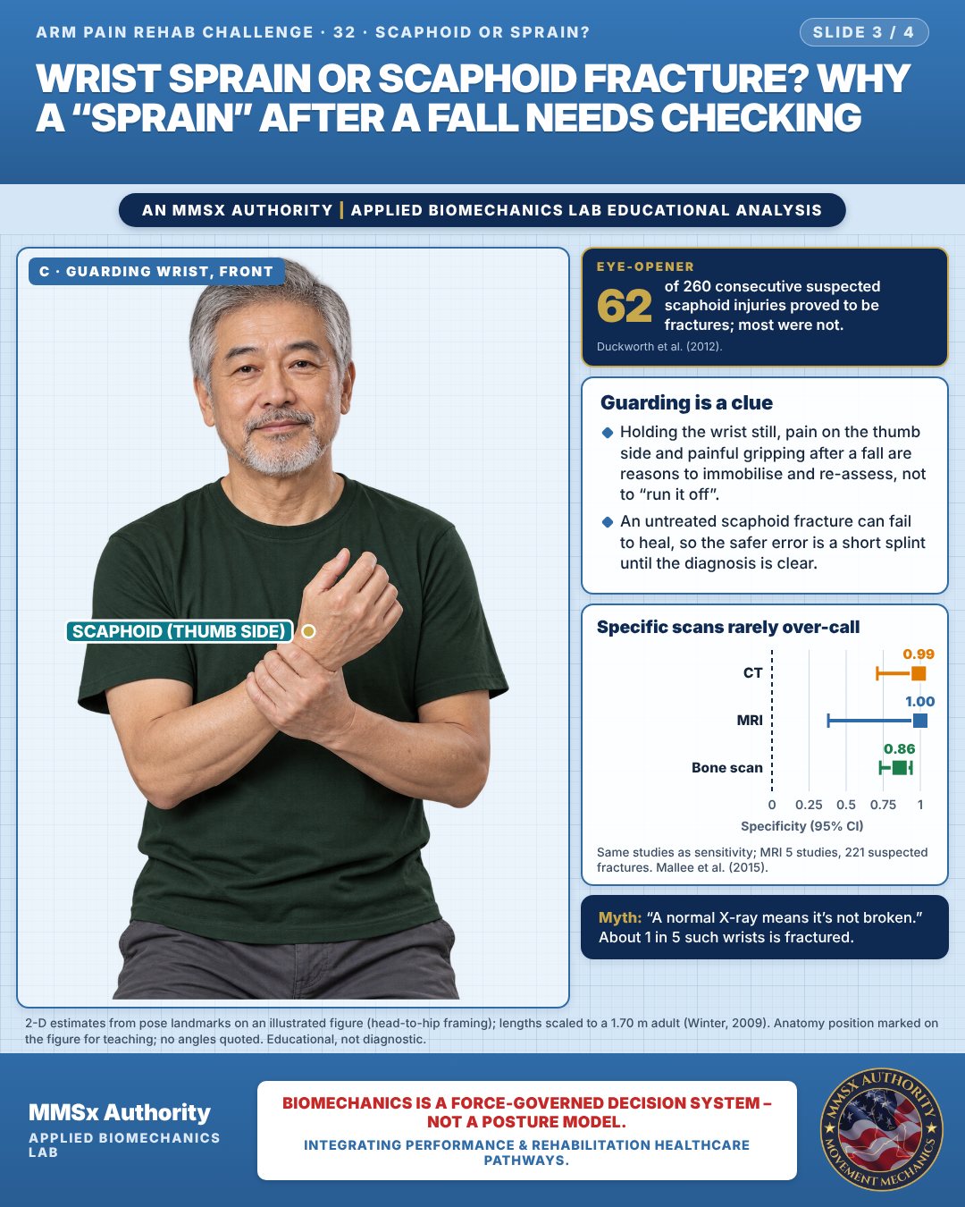
Guarding is a clue
Holding the wrist still, pain on the thumb side and painful gripping after a fall are reasons to immobilise and re-assess, not to “run it off”.
An untreated scaphoid fracture can fail to heal, so the safer error is a short splint until the diagnosis is clear.
Specific scans rarely over-call
Decision pathway
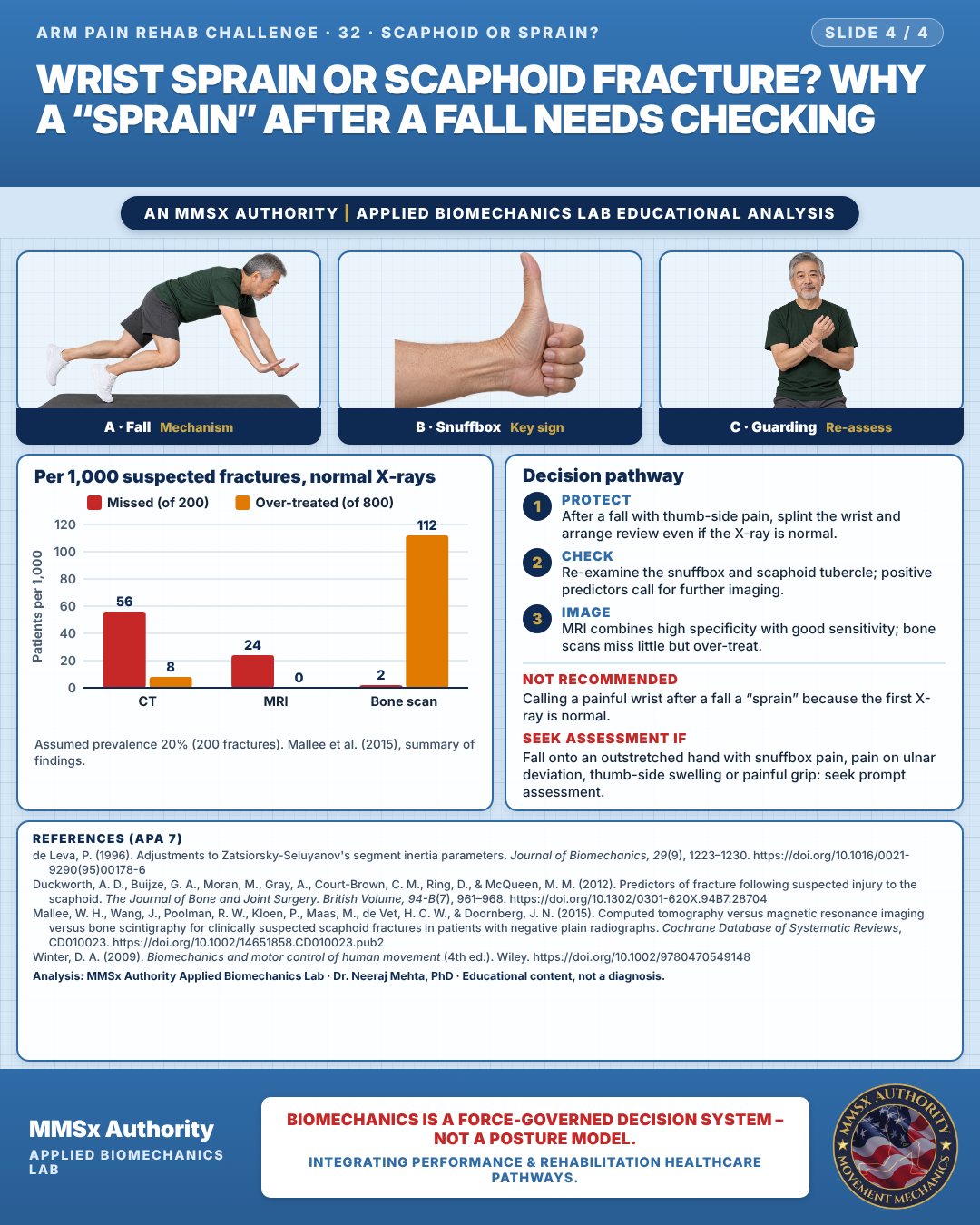
Per 1,000 suspected fractures, normal X-rays
Protect
After a fall with thumb-side pain, splint the wrist and arrange review even if the X-ray is normal.
Check
Re-examine the snuffbox and scaphoid tubercle; positive predictors call for further imaging.
Image
MRI combines high specificity with good sensitivity; bone scans miss little but over-treat.
Myth and evidence
Myth“A normal X-ray means it's not broken.”
EvidenceAmong suspected fractures with normal initial X-rays, about 1 in 5 (median 20%) are true fractures (Mallee et al., 2015).
Cautions
Not recommended
Calling a painful wrist after a fall a “sprain” because the first X-ray is normal.
Seek assessment if
Fall onto an outstretched hand with snuffbox pain, pain on ulnar deviation, thumb-side swelling or painful grip: seek prompt assessment.
Methods note
2-D estimates from pose landmarks on an illustrated figure; CoM (if shown) from de Leva (1996); cm for a 1.70 m adult (Winter, 2009). Wrist angle from manually placed forearm and hand axes. Angles from landmarks placed on an illustrated figure (2-D, camera-plane); approximate. Forearm axis and anatomy positions marked on the figure for teaching. 2-D estimates from pose landmarks on an illustrated figure (head-to-hip framing); lengths scaled to a 1.70 m adult (Winter, 2009). Anatomy position marked on the figure for teaching; no angles quoted. These are educational estimates, not a diagnosis.
References
- de Leva, P. (1996). Adjustments to Zatsiorsky-Seluyanov's segment inertia parameters. Journal of Biomechanics, 29(9), 1223–1230. https://doi.org/10.1016/0021-9290(95)00178-6
- Duckworth, A. D., Buijze, G. A., Moran, M., Gray, A., Court-Brown, C. M., Ring, D., & McQueen, M. M. (2012). Predictors of fracture following suspected injury to the scaphoid. The Journal of Bone and Joint Surgery. British Volume, 94-B(7), 961–968. https://doi.org/10.1302/0301-620X.94B7.28704
- Mallee, W. H., Wang, J., Poolman, R. W., Kloen, P., Maas, M., de Vet, H. C. W., & Doornberg, J. N. (2015). Computed tomography versus magnetic resonance imaging versus bone scintigraphy for clinically suspected scaphoid fractures in patients with negative plain radiographs. Cochrane Database of Systematic Reviews, CD010023. https://doi.org/10.1002/14651858.CD010023.pub2
- Winter, D. A. (2009). Biomechanics and motor control of human movement (4th ed.). Wiley. https://doi.org/10.1002/9780470549148
Cite this article
Mehta, N. (2026, October 28). Wrist sprain or scaphoid fracture? Why a “sprain” after a fall needs checking. MMSx Authority Applied Biomechanics Lab. https://mmsxauthority.org/edu/analysis/arm/32-wrist-sprain-or-scaphoid-fracture-why-a-sprain-after-a-fall-needs/
Part of the collection
Mehta, N. (2026). Elbow, Forearm, Wrist and Hand Rehabilitation: 50 Evidence-Based Biomechanics Analyses [Collection]. MMSx Authority Applied Biomechanics Lab. Zenodo. https://doi.org/10.5281/zenodo.23735910
All Arm Pain Rehab Challenge articles · Rehabilitation Library