Arm Pain Rehab Challenge · 18
Cubital tunnel syndrome: tingling in the ring and little fingers
Compression or traction neuropathy of the ulnar nerve at the elbow (ulnar neuropathy at the elbow, UNE), typically producing numbness or tingling in…
Key points
- Modify. Use a headset or speaker, stop leaning on the elbow and break up long periods of deep elbow bending.
- Advise. Explain the nerve’s path and which positions provoke it; simple information did as well as splints or gliding.
- Escalate. Persisting symptoms or weakness: nerve-conduction testing; if operating, simple decompression is preferred.
What it is
Compression or traction neuropathy of the ulnar nerve at the elbow (ulnar neuropathy at the elbow, UNE), typically producing numbness or tingling in the little finger and the little-finger half of the ring finger, and in more advanced cases weakness of the intrinsic hand muscles.
How we analysed the movement
Three movements relevant to this topic were analysed from illustrated reference figures. Joint centres were located with two-dimensional pose estimation where the whole arm was visible; elbow and wrist angles, and the directions of external load and tendon pull, were then drawn on the figure; on hand close-ups, where pose estimation does not apply, the landmarks were placed manually. The figures are educational estimates that show how position changes mechanical demand; they are not measurements of a patient. Every research value quoted below comes from the published source cited beside it.
Movement A: Phone to ear (side view)
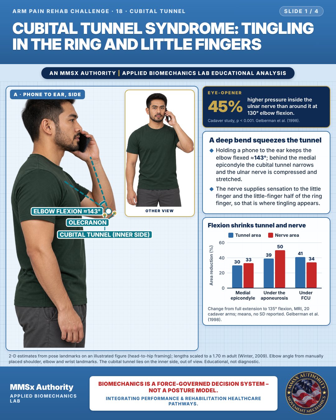
A deep bend squeezes the tunnel
Holding a phone to the ear keeps the elbow flexed ≈143°; behind the medial epicondyle the cubital tunnel narrows and the ulnar nerve is compressed and stretched.
The nerve supplies sensation to the little finger and the little-finger half of the ring finger, so that is where tingling appears.
Flexion shrinks tunnel and nerve
Movement B: Palm up, top
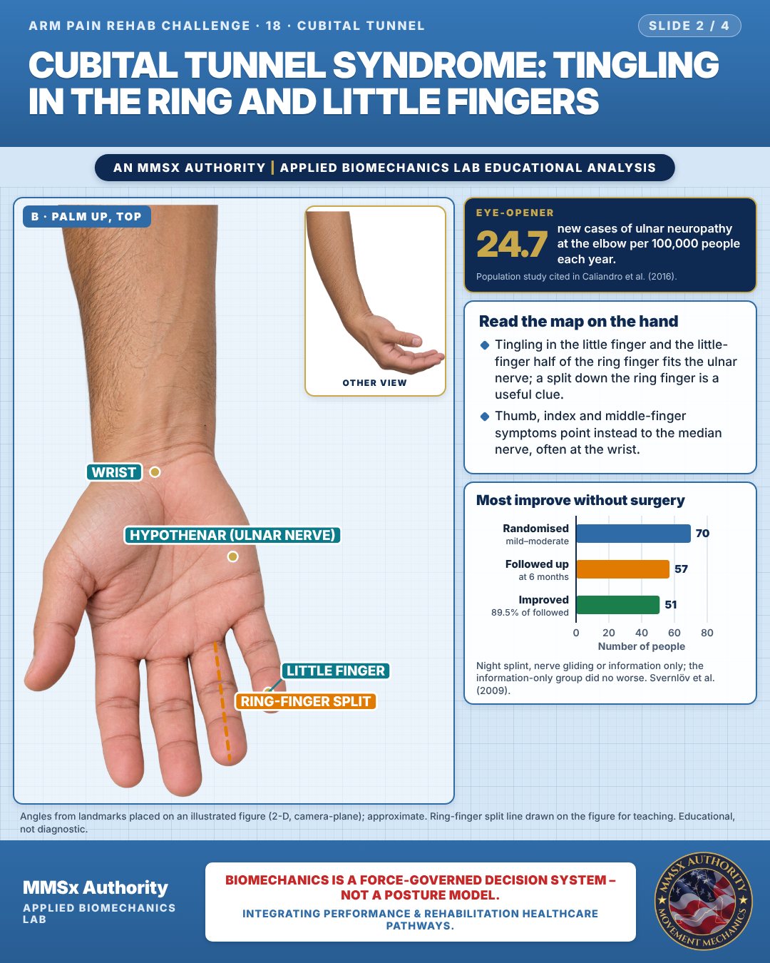
Read the map on the hand
Tingling in the little finger and the little-finger half of the ring finger fits the ulnar nerve; a split down the ring finger is a useful clue.
Thumb, index and middle-finger symptoms point instead to the median nerve, often at the wrist.
Most improve without surgery
Movement C: Elbow on desk (side view)
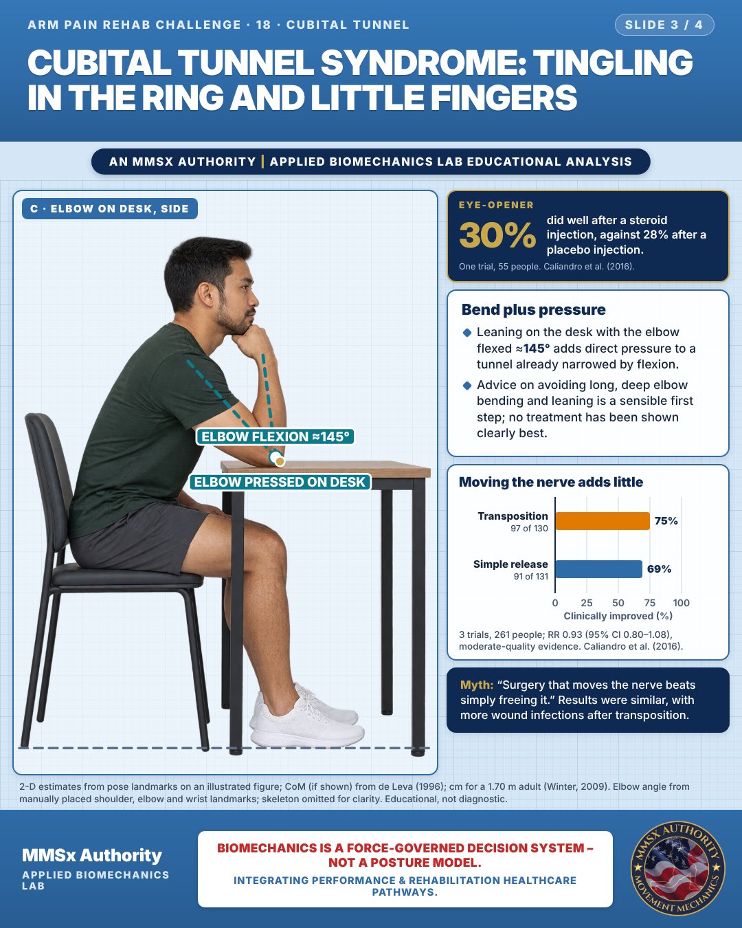
Bend plus pressure
Leaning on the desk with the elbow flexed ≈145° adds direct pressure to a tunnel already narrowed by flexion.
Advice on avoiding long, deep elbow bending and leaning is a sensible first step; no treatment has been shown clearly best.
Moving the nerve adds little
Decision pathway
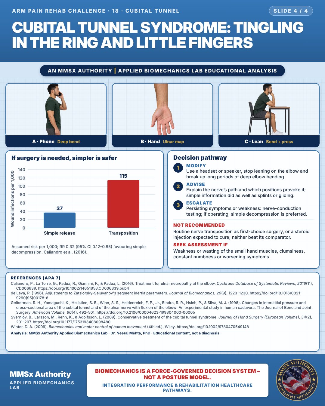
If surgery is needed, simpler is safer
Modify
Use a headset or speaker, stop leaning on the elbow and break up long periods of deep elbow bending.
Advise
Explain the nerve’s path and which positions provoke it; simple information did as well as splints or gliding.
Escalate
Persisting symptoms or weakness: nerve-conduction testing; if operating, simple decompression is preferred.
What the evidence supports
- Evidence insufficient to identify the best treatment from clinical, neurophysiological and imaging features. For mild symptoms, information on movements/positions to avoid may reduce discomfort. When surgery is chosen, simple decompression and transposition give similar clinical results; simple decompression has fewer wound infections. Steroid injection was not better than placebo in the one trial (Caliandro et al., 2016).
Myth and evidence
Myth“Surgery that moves the nerve is always better than freeing it.”
EvidenceModerate-quality evidence shows no difference in clinical improvement (RR 0.93, 95% CI 0.80–1.08) but more wound infections with transposition (Caliandro et al., 2016).
Cautions
Not recommended
Routine nerve transposition as first-choice surgery, or a steroid injection expected to cure; neither beat its comparator.
Seek assessment if
Weakness or wasting of the small hand muscles, clumsiness, constant numbness or worsening symptoms.
Methods note
2-D estimates from pose landmarks on an illustrated figure (head-to-hip framing); lengths scaled to a 1.70 m adult (Winter, 2009). Elbow angle from manually placed shoulder, elbow and wrist landmarks. The cubital tunnel lies on the inner side, out of view. Angles from landmarks placed on an illustrated figure (2-D, camera-plane); approximate. Ring-finger split line drawn on the figure for teaching. 2-D estimates from pose landmarks on an illustrated figure; CoM (if shown) from de Leva (1996); cm for a 1.70 m adult (Winter, 2009). Elbow angle from manually placed shoulder, elbow and wrist landmarks; skeleton omitted for clarity. These are educational estimates, not a diagnosis.
References
- Caliandro, P., La Torre, G., Padua, R., Giannini, F., & Padua, L. (2016). Treatment for ulnar neuropathy at the elbow. Cochrane Database of Systematic Reviews, 2016(11), CD006839. https://doi.org/10.1002/14651858.CD006839.pub4
- de Leva, P. (1996). Adjustments to Zatsiorsky-Seluyanov's segment inertia parameters. Journal of Biomechanics, 29(9), 1223–1230. https://doi.org/10.1016/0021-9290(95)00178-6
- Gelberman, R. H., Yamaguchi, K., Hollstien, S. B., Winn, S. S., Heidenreich, F. P., Jr., Bindra, R. R., Hsieh, P., & Silva, M. J. (1998). Changes in interstitial pressure and cross-sectional area of the cubital tunnel and of the ulnar nerve with flexion of the elbow: An experimental study in human cadavera. The Journal of Bone and Joint Surgery. American Volume, 80(4), 492–501. https://doi.org/10.2106/00004623-199804000-00005
- Svernlöv, B., Larsson, M., Rehn, K., & Adolfsson, L. (2009). Conservative treatment of the cubital tunnel syndrome. Journal of Hand Surgery (European Volume), 34(2), 201–207. https://doi.org/10.1177/1753193408098480
- Winter, D. A. (2009). Biomechanics and motor control of human movement (4th ed.). Wiley. https://doi.org/10.1002/9780470549148
Cite this article
Mehta, N. (2026, October 28). Cubital tunnel syndrome: tingling in the ring and little fingers. MMSx Authority Applied Biomechanics Lab. https://mmsxauthority.org/edu/analysis/arm/18-cubital-tunnel-syndrome-tingling-in-the-ring-and-little-fingers/
Part of the collection
Mehta, N. (2026). Elbow, Forearm, Wrist and Hand Rehabilitation: 50 Evidence-Based Biomechanics Analyses [Collection]. MMSx Authority Applied Biomechanics Lab. Zenodo. https://doi.org/10.5281/zenodo.23735910
All Arm Pain Rehab Challenge articles · Rehabilitation Library