Lower Back Rehab Challenge · 50
Exercise after lumbar disc surgery: why progression must be individualised
Rehabilitation after lumbar disc surgery (usually discectomy or microdiscectomy for a herniated disc causing sciatica) is a structured, progressive…
Key points
- Coordinate. Follow the surgeon’s advice on timing; agree the plan with the surgical team.
- Start at 4–6 weeks. Structured exercise usually begins then; walking and daily activity earlier as advised.
- Progress individually. Increase intensity by symptoms and function; no one-size-fits-all timetable.
What it is
Rehabilitation after lumbar disc surgery (usually discectomy or microdiscectomy for a herniated disc causing sciatica) is a structured, progressive programme of activity and exercise — walking, trunk and hip strengthening, flexibility and return to work/sport tasks — usually starting from about 4–6 weeks after surgery in trials, adapted to the procedure, surgeon's advice, wound healing, and the person's symptoms and goals.
How we analysed the movement
Three movements relevant to this topic were analysed from illustrated reference figures. Joint centres were located with two-dimensional pose estimation; joint angles, the whole-body centre of mass and, where relevant, the low-back lever (the horizontal distance from the upper-body centre of mass to the L5/S1 joint) were then calculated from standard segment parameters. The figures are educational estimates that show how position changes mechanical demand; they are not measurements of a patient. Every research value quoted below comes from the published source cited beside it.
Movement A: Individual plan, seated
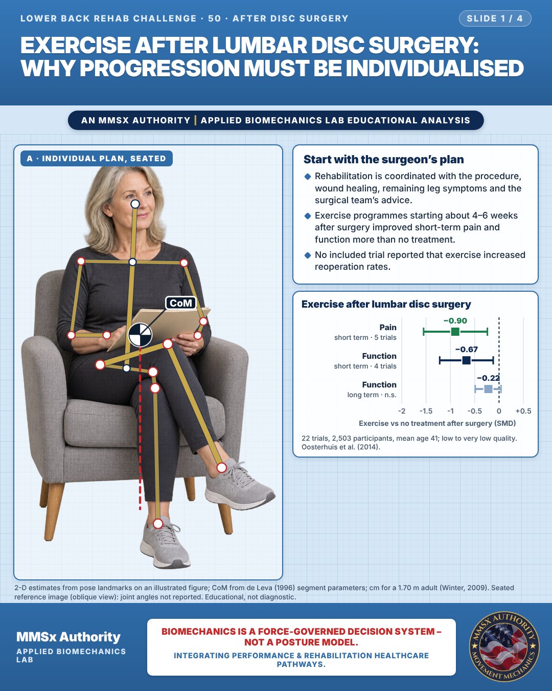
Start with the surgeon’s plan
Rehabilitation is coordinated with the procedure, wound healing, remaining leg symptoms and the surgical team’s advice.
Exercise programmes starting about 4–6 weeks after surgery improved short-term pain and function more than no treatment.
No included trial reported that exercise increased reoperation rates.
Exercise after lumbar disc surgery
Movement B: Supported walk (front view)
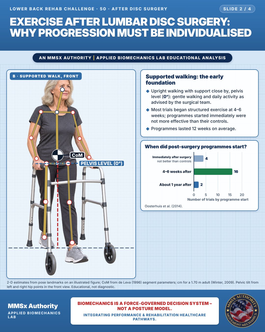
Supported walking: the early foundation
Upright walking with support close by, pelvis level (0°): gentle walking and daily activity as advised by the surgical team.
Most trials began structured exercise at 4–6 weeks; programmes started immediately were not more effective than their controls.
Programmes lasted 12 weeks on average.
When did post-surgery programmes start?
Movement C: Supported transfer
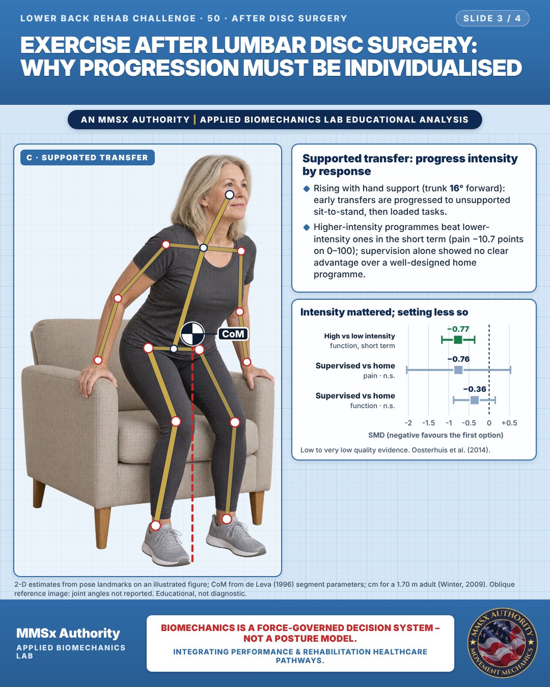
Supported transfer: progress intensity by response
Rising with hand support (trunk 16° forward): early transfers are progressed to unsupported sit-to-stand, then loaded tasks.
Higher-intensity programmes beat lower-intensity ones in the short term (pain −10.7 points on 0–100); supervision alone showed no clear advantage over a well-designed home programme.
Intensity mattered; setting less so
Decision pathway
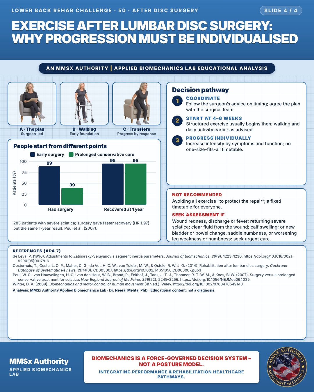
People start from different points
Coordinate
Follow the surgeon’s advice on timing; agree the plan with the surgical team.
Start at 4–6 weeks
Structured exercise usually begins then; walking and daily activity earlier as advised.
Progress individually
Increase intensity by symptoms and function; no one-size-fits-all timetable.
What the evidence supports
- Recommend: follow the surgeon's specific post-operative advice; early gentle walking and daily activity as advised; a progressive exercise programme typically from about 4–6 weeks, progressed in intensity according to symptoms and function (Oosterhuis et al., 2014); home-based programmes can be appropriate when well designed (Oosterhuis et al., 2014).
- Do not: use a one-size-fits-all timetable; assume exercise increases the risk of reoperation (no included trial reported this) (Oosterhuis et al., 2014); ignore new or returning leg symptoms.
Cautions
Not recommended
Avoiding all exercise “to protect the repair”; a fixed timetable for everyone.
Seek assessment if
Wound redness, discharge or fever; returning severe sciatica; clear fluid from the wound; calf swelling; or new bladder or bowel change, saddle numbness, or worsening leg weakness or numbness: seek urgent care.
Methods note
2-D estimates from pose landmarks on an illustrated figure; CoM from de Leva (1996) segment parameters; cm for a 1.70 m adult (Winter, 2009). Seated reference image (oblique view): joint angles not reported. Pelvic tilt from left and right hip points in the front view. Oblique reference image: joint angles not reported. These are educational estimates, not a diagnosis.
References
- de Leva, P. (1996). Adjustments to Zatsiorsky-Seluyanov's segment inertia parameters. Journal of Biomechanics, 29(9), 1223–1230. https://doi.org/10.1016/0021-9290(95)00178-6
- Oosterhuis, T., Costa, L. O. P., Maher, C. G., de Vet, H. C. W., van Tulder, M. W., & Ostelo, R. W. J. G. (2014). Rehabilitation after lumbar disc surgery. Cochrane Database of Systematic Reviews, 2014(3), CD003007. https://doi.org/10.1002/14651858.CD003007.pub3
- Peul, W. C., van Houwelingen, H. C., van den Hout, W. B., Brand, R., Eekhof, J., Tans, J. T. J., Thomeer, R. T. W. M., & Koes, B. W. (2007). Surgery versus prolonged conservative treatment for sciatica. New England Journal of Medicine, 356(22), 2245–2256. https://doi.org/10.1056/NEJMoa064039
- Winter, D. A. (2009). Biomechanics and motor control of human movement (4th ed.). Wiley. https://doi.org/10.1002/9780470549148
Cite this article
Mehta, N. (2026, October 23). Exercise after lumbar disc surgery: why progression must be individualised. MMSx Authority Applied Biomechanics Lab. https://mmsxauthority.org/edu/analysis/lower-back/50-exercise-after-lumbar-disc-surgery-why-progression-must-be/
Part of the collection
Mehta, N. (2026). Lower Back Rehabilitation: 50 Evidence-Based Biomechanics Analyses [Collection]. MMSx Authority Applied Biomechanics Lab. Zenodo. https://doi.org/10.5281/zenodo.23735778
All Lower Back Rehab Challenge articles · Rehabilitation Library