Knee Rehab Challenge · 13
Why do my knees hurt when I squat?
Knee pain with squatting is a symptom, not a diagnosis: it most commonly reflects patellofemoral pain (pain around/behind the kneecap with loaded…
Key points
- Dose depth. Start in a comfortable range (about 0–50° when load must be minimised), then progress depth.
- Dose load. Two legs before one, bodyweight before barbell; knee travel is one dial among depth and load.
- Build. Hip- and knee-targeted exercise is grade A for patellofemoral pain; squats are a usable therapeutic exercise.
What it is
Knee pain with squatting is a symptom, not a diagnosis: it most commonly reflects patellofemoral pain (pain around/behind the kneecap with loaded knee flexion) or knee osteoarthritis, and squat load on the knee rises with depth, external load and forward knee travel (Willy et al., 2019; Wallace et al., 2002).
How we analysed the movement
Three movements relevant to this topic were analysed from illustrated reference figures. Joint centres were located with two-dimensional pose estimation; joint angles, the whole-body centre of mass and, where relevant, the ground-reaction-force line and its moment arm about the knee were then calculated from standard segment parameters. The figures are educational estimates that show how position changes mechanical demand; they are not measurements of a patient. Every research value quoted below comes from the published source cited beside it.
Movement A: Bodyweight squat (side view)
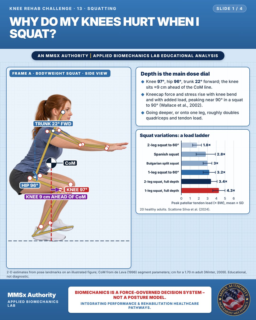
Depth is the main dose dial
Knee 97°, hip 96°, trunk 22° forward; the knee sits ≈9 cm ahead of the CoM line.
Kneecap force and stress rise with knee bend and with added load, peaking near 90° in a squat to 90° (Wallace et al., 2002).
Going deeper, or onto one leg, roughly doubles quadriceps and tendon load.
Squat variations: a load ladder
Movement B: Bodyweight squat (front view)
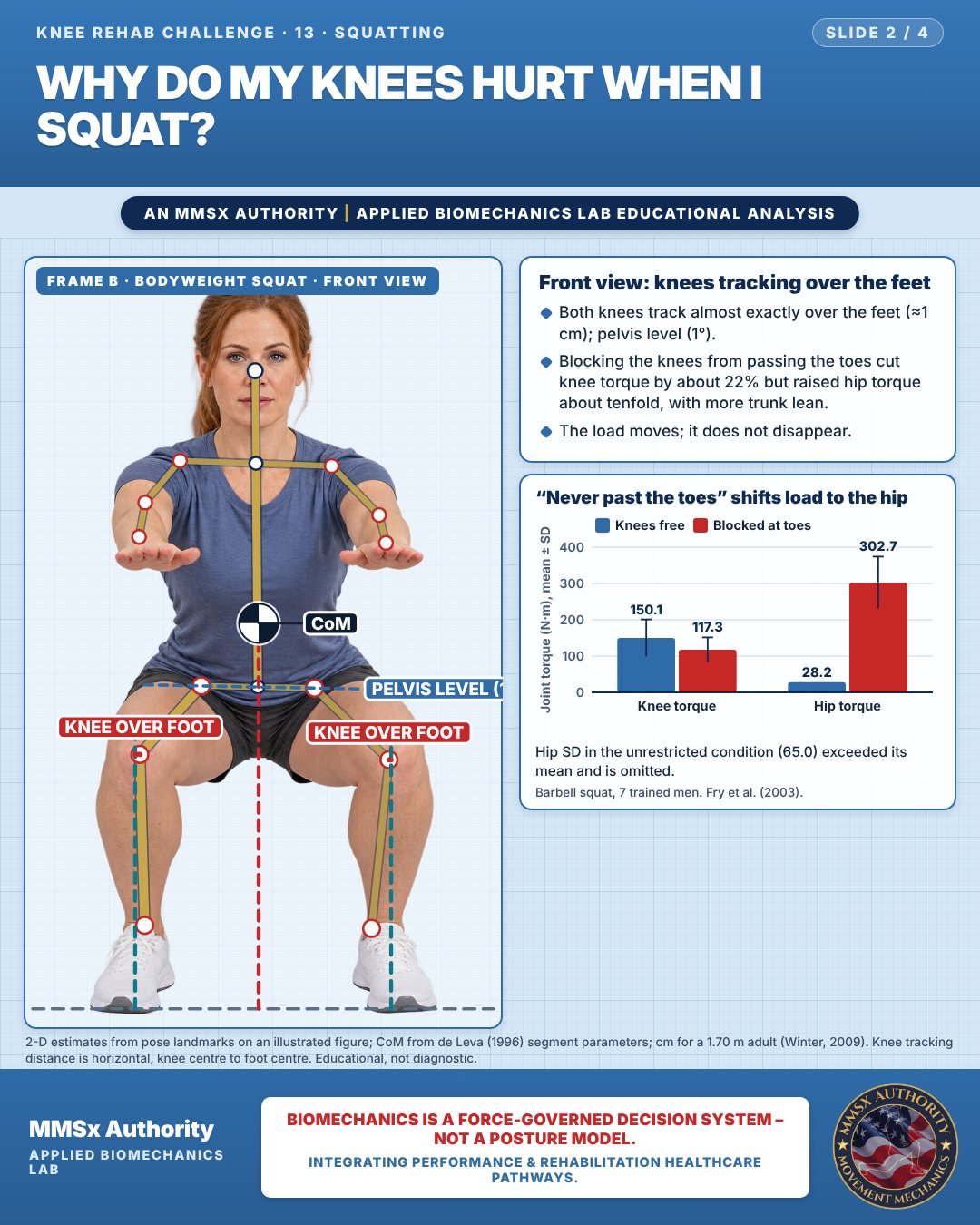
Front view: knees tracking over the feet
Both knees track almost exactly over the feet (≈1 cm); pelvis level (1°).
Blocking the knees from passing the toes cut knee torque by about 22% but raised hip torque about tenfold, with more trunk lean.
The load moves; it does not disappear.
“Never past the toes” shifts load to the hip
Hip SD in the unrestricted condition (65.0) exceeded its mean and is omitted.
Movement C: Lifting a box (side view)
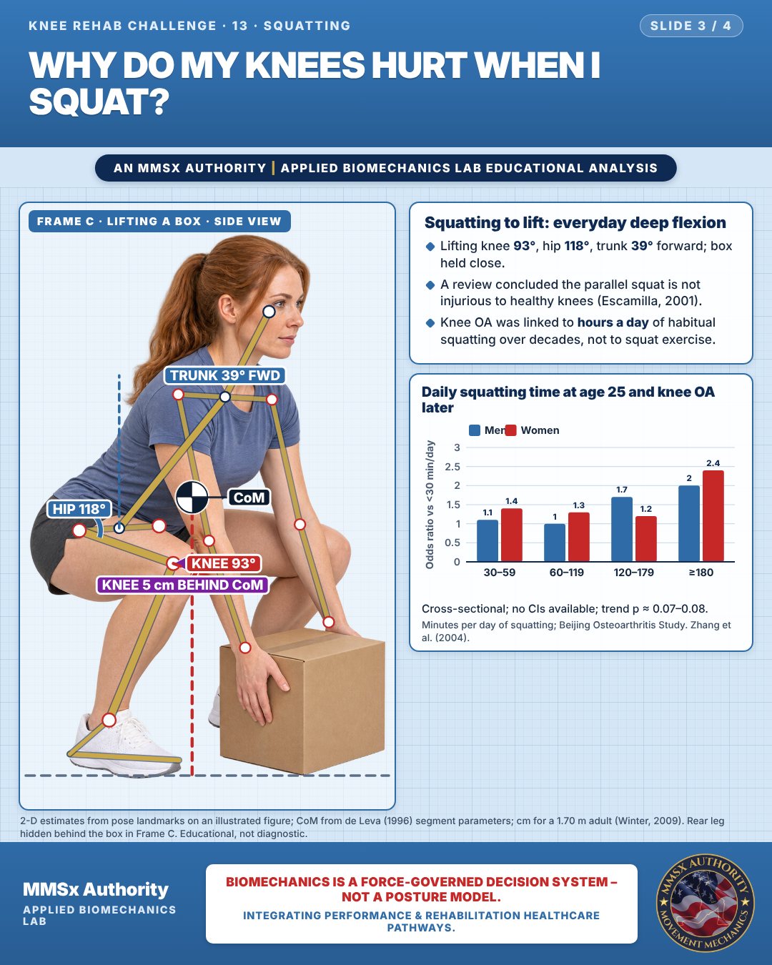
Squatting to lift: everyday deep flexion
Lifting knee 93°, hip 118°, trunk 39° forward; box held close.
A review concluded the parallel squat is not injurious to healthy knees (Escamilla, 2001).
Knee OA was linked to hours a day of habitual squatting over decades, not to squat exercise.
Daily squatting time at age 25 and knee OA later
Cross-sectional; no CIs available; trend p ≈ 0.07–0.08.
Decision pathway
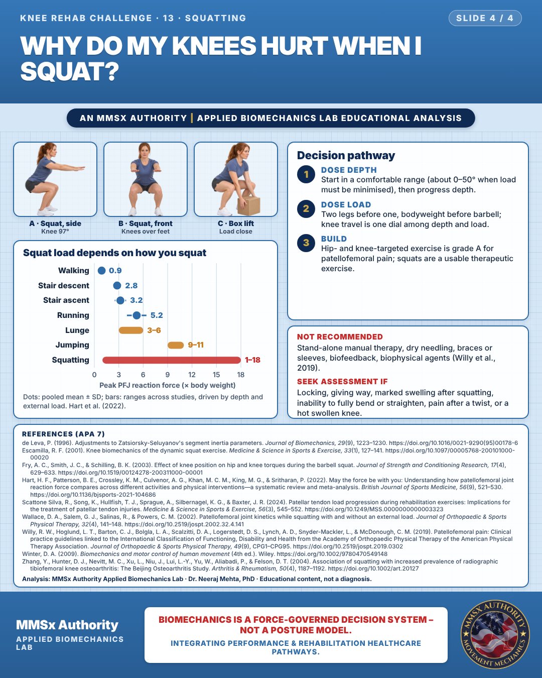
Squat load depends on how you squat
Dose depth
Start in a comfortable range (about 0–50° when load must be minimised), then progress depth.
Dose load
Two legs before one, bodyweight before barbell; knee travel is one dial among depth and load.
Build
Hip- and knee-targeted exercise is grade A for patellofemoral pain; squats are a usable therapeutic exercise.
What the evidence supports
- Not recommended: stand-alone manual therapy, dry needling, braces/sleeves, biofeedback, biophysical agents (Willy et al., 2019). Avoid blanket bans on deep squats or on knees passing the toes in healthy knees (Escamilla, 2001; Fry et al., 2003).
Myth and evidence
Myth“Never let your knees go past your toes.”
EvidenceBlocking forward knee travel cut knee torque by ~22% but raised hip torque from 28 to 303 N·m (Fry et al., 2003) — the load moves, it does not disappear. Accurate framing: knees-forward squats do raise patellofemoral load (Hart et al., 2022), so for an irritable patellofemoral joint, knee travel is one dose dial among depth and external load — not a universal rule.
Myth“Deep squats wreck healthy knees.”
EvidenceThe dynamic-squat review concluded the parallel squat is not injurious to healthy knees (Escamilla, 2001); load rises with depth (Wallace et al., 2002; Scattone Silva et al., 2024), so depth is a dose to be progressed, not forbidden. The OA association is with many hours a day of habitual squatting over decades (Zhang et al., 2004).
Cautions
Not recommended
Stand-alone manual therapy, dry needling, braces or sleeves, biofeedback, biophysical agents (Willy et al., 2019).
Seek assessment if
Locking, giving way, marked swelling after squatting, inability to fully bend or straighten, pain after a twist, or a hot swollen knee.
Methods note
2-D estimates from pose landmarks on an illustrated figure; CoM from de Leva (1996) segment parameters; cm for a 1.70 m adult (Winter, 2009). Knee tracking distance is horizontal, knee centre to foot centre. Rear leg hidden behind the box in Frame C. These are educational estimates, not a diagnosis.
References
- de Leva, P. (1996). Adjustments to Zatsiorsky-Seluyanov's segment inertia parameters. Journal of Biomechanics, 29(9), 1223–1230. https://doi.org/10.1016/0021-9290(95)00178-6
- Escamilla, R. F. (2001). Knee biomechanics of the dynamic squat exercise. Medicine & Science in Sports & Exercise, 33(1), 127–141. https://doi.org/10.1097/00005768-200101000-00020
- Fry, A. C., Smith, J. C., & Schilling, B. K. (2003). Effect of knee position on hip and knee torques during the barbell squat. Journal of Strength and Conditioning Research, 17(4), 629–633. https://doi.org/10.1519/00124278-200311000-00001
- Hart, H. F., Patterson, B. E., Crossley, K. M., Culvenor, A. G., Khan, M. C. M., King, M. G., & Sritharan, P. (2022). May the force be with you: Understanding how patellofemoral joint reaction force compares across different activities and physical interventions—a systematic review and meta-analysis. British Journal of Sports Medicine, 56(9), 521–530. https://doi.org/10.1136/bjsports-2021-104686
- Scattone Silva, R., Song, K., Hullfish, T. J., Sprague, A., Silbernagel, K. G., & Baxter, J. R. (2024). Patellar tendon load progression during rehabilitation exercises: Implications for the treatment of patellar tendon injuries. Medicine & Science in Sports & Exercise, 56(3), 545–552. https://doi.org/10.1249/MSS.0000000000003323
- Wallace, D. A., Salem, G. J., Salinas, R., & Powers, C. M. (2002). Patellofemoral joint kinetics while squatting with and without an external load. Journal of Orthopaedic & Sports Physical Therapy, 32(4), 141–148. https://doi.org/10.2519/jospt.2002.32.4.141
- Willy, R. W., Hoglund, L. T., Barton, C. J., Bolgla, L. A., Scalzitti, D. A., Logerstedt, D. S., Lynch, A. D., Snyder-Mackler, L., & McDonough, C. M. (2019). Patellofemoral pain: Clinical practice guidelines linked to the International Classification of Functioning, Disability and Health from the Academy of Orthopaedic Physical Therapy of the American Physical Therapy Association. Journal of Orthopaedic & Sports Physical Therapy, 49(9), CPG1–CPG95. https://doi.org/10.2519/jospt.2019.0302
- Winter, D. A. (2009). Biomechanics and motor control of human movement (4th ed.). Wiley. https://doi.org/10.1002/9780470549148
- Zhang, Y., Hunter, D. J., Nevitt, M. C., Xu, L., Niu, J., Lui, L.-Y., Yu, W., Aliabadi, P., & Felson, D. T. (2004). Association of squatting with increased prevalence of radiographic tibiofemoral knee osteoarthritis: The Beijing Osteoarthritis Study. Arthritis & Rheumatism, 50(4), 1187–1192. https://doi.org/10.1002/art.20127
Cite this article
Mehta, N. (2026, October 23). Why do my knees hurt when I squat? MMSx Authority Applied Biomechanics Lab. https://mmsxauthority.org/edu/analysis/knee/13-why-do-my-knees-hurt-when-i-squat/
Part of the collection
Mehta, N. (2026). Knee Rehabilitation: 50 Evidence-Based Biomechanics Analyses [Collection]. MMSx Authority Applied Biomechanics Lab. Zenodo. https://doi.org/10.5281/zenodo.23735752