Hip & Groin Rehab Challenge · 19
Hip abductor strength: the exercise evidence for hip pain
Hip abductor strengthening is progressive resistance training of gluteus medius and minimus (with tensor fasciae latae), using side-lying, standing,…
Key points
- Diagnose. Separate gluteal tendinopathy from hip osteoarthritis; the evidence for abductor exercise differs between them.
- Progress. Move from step-ups and side-lying work to single-limb squats and side planks as pain allows.
- Combine. Pair exercise with education; in hip OA place abductor work inside a broader, individual plan.
What it is
Hip abductor strengthening is progressive resistance training of gluteus medius and minimus (with tensor fasciae latae), using side-lying, standing, banded and single-leg exercises. It is a core part of exercise programmes for lateral hip pain (gluteal tendinopathy) and is also used in hip osteoarthritis.
How we analysed the movement
Three movements relevant to this topic were analysed from illustrated reference figures. Joint centres were located with two-dimensional pose estimation; hip and knee angles, pelvis level, the whole-body centre of mass and, where relevant, the hip lever (the horizontal distance from the hip joint to the upper-body centre of mass) and the resulting static hip moment were then calculated from standard segment parameters. The figures are educational estimates that show how position changes mechanical demand; they are not measurements of a patient. Every research value quoted below comes from the published source cited beside it.
Movement A: Side plank (front view)
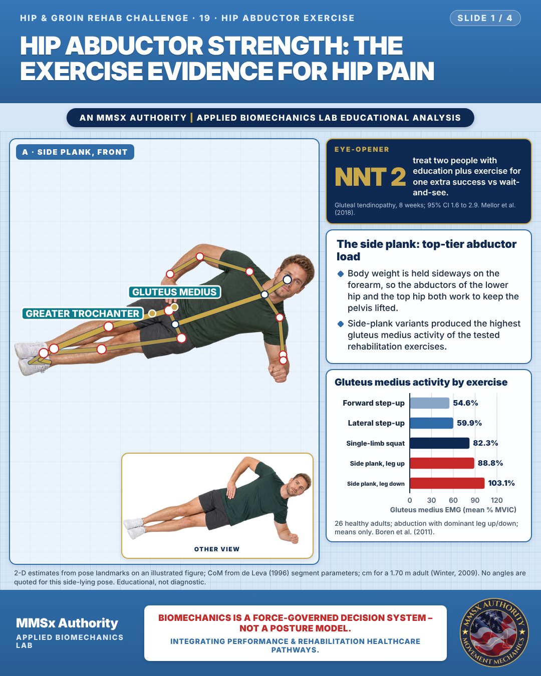
The side plank: top-tier abductor load
Body weight is held sideways on the forearm, so the abductors of the lower hip and the top hip both work to keep the pelvis lifted.
Side-plank variants produced the highest gluteus medius activity of the tested rehabilitation exercises.
Gluteus medius activity by exercise
Movement B: Banded side step (front view)
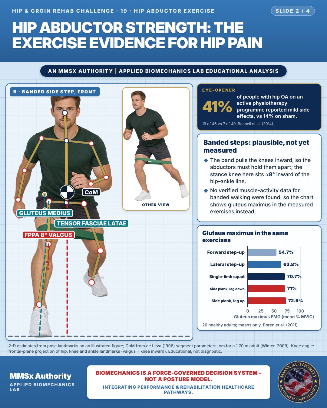
Banded steps: plausible, not yet measured
The band pulls the knees inward, so the abductors must hold them apart; the stance knee here sits ≈8° inward of the hip–ankle line.
No verified muscle-activity data for banded walking were found, so the chart shows gluteus maximus in the measured exercises instead.
Gluteus maximus in the same exercises
Movement C: Step-down (front view)
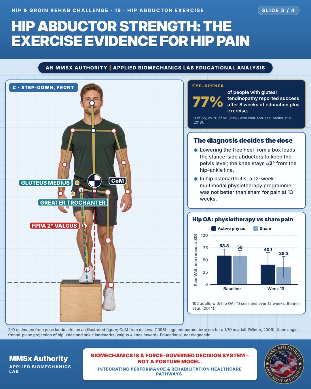
The diagnosis decides the dose
Lowering the free heel from a box loads the stance-side abductors to keep the pelvis level; the knee stays ≈2° from the hip–ankle line.
In hip osteoarthritis, a 12-week multimodal physiotherapy programme was not better than sham for pain at 13 weeks.
Hip OA: physiotherapy vs sham pain
Decision pathway
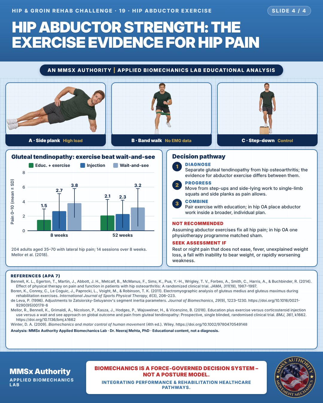
Gluteal tendinopathy: exercise beat wait-and-see
Diagnose
Separate gluteal tendinopathy from hip osteoarthritis; the evidence for abductor exercise differs between them.
Progress
Move from step-ups and side-lying work to single-limb squats and side planks as pain allows.
Combine
Pair exercise with education; in hip OA place abductor work inside a broader, individual plan.
What the evidence supports
- Gluteal tendinopathy: education plus progressive exercise is recommended over injection and wait-and-see (Mellor et al., 2018).
- Hip osteoarthritis: one trial of a multimodal physiotherapy programme found no advantage over sham at 13 weeks, with more mild adverse effects (Bennell et al., 2014); abductor training should sit within a broader, individualised plan.
- Exercise choice: progress from lower-activation (step-ups, side-lying) to higher-activation exercises (single-limb squat, side plank) (Boren et al., 2011).
Myth and evidence
Myth“Hip abductor exercises fix all hip pain.”
EvidenceThey worked well for gluteal tendinopathy (Mellor et al., 2018), but a multimodal physiotherapy programme was not better than sham for hip osteoarthritis pain at 13 weeks (Bennell et al., 2014). The diagnosis decides the dose.
Cautions
Not recommended
Assuming abductor exercises fix all hip pain; in hip OA one physiotherapy programme matched sham.
Seek assessment if
Rest or night pain that does not ease, fever, unexplained weight loss, a fall with inability to bear weight, or rapidly worsening weakness.
Methods note
2-D estimates from pose landmarks on an illustrated figure; CoM from de Leva (1996) segment parameters; cm for a 1.70 m adult (Winter, 2009). No angles are quoted for this side-lying pose. Knee angle: frontal-plane projection of hip, knee and ankle landmarks (valgus = knee inward). These are educational estimates, not a diagnosis.
References
- Bennell, K. L., Egerton, T., Martin, J., Abbott, J. H., Metcalf, B., McManus, F., Sims, K., Pua, Y.-H., Wrigley, T. V., Forbes, A., Smith, C., Harris, A., & Buchbinder, R. (2014). Effect of physical therapy on pain and function in patients with hip osteoarthritis: A randomized clinical trial. JAMA, 311(19), 1987–1997.
- Boren, K., Conrey, C., Le Coguic, J., Paprocki, L., Voight, M., & Robinson, T. K. (2011). Electromyographic analysis of gluteus medius and gluteus maximus during rehabilitation exercises. International Journal of Sports Physical Therapy, 6(3), 206–223.
- de Leva, P. (1996). Adjustments to Zatsiorsky-Seluyanov's segment inertia parameters. Journal of Biomechanics, 29(9), 1223–1230. https://doi.org/10.1016/0021-9290(95)00178-6
- Mellor, R., Bennell, K., Grimaldi, A., Nicolson, P., Kasza, J., Hodges, P., Wajswelner, H., & Vicenzino, B. (2018). Education plus exercise versus corticosteroid injection use versus a wait and see approach on global outcome and pain from gluteal tendinopathy: Prospective, single blinded, randomised clinical trial. BMJ, 361, k1662. https://doi.org/10.1136/bmj.k1662
- Winter, D. A. (2009). Biomechanics and motor control of human movement (4th ed.). Wiley. https://doi.org/10.1002/9780470549148
Cite this article
Mehta, N. (2026, October 28). Hip abductor strength: the exercise evidence for hip pain. MMSx Authority Applied Biomechanics Lab. https://mmsxauthority.org/edu/analysis/hip-groin/19-hip-abductor-strength-the-exercise-evidence-for-hip-pain/
Part of the collection
Mehta, N. (2026). Hip and Groin Rehabilitation: 30 Evidence-Based Biomechanics Analyses [Collection]. MMSx Authority Applied Biomechanics Lab. Zenodo. https://doi.org/10.5281/zenodo.23735806
All Hip & Groin Rehab Challenge articles · Rehabilitation Library