Hip & Groin Rehab Challenge · 17
Glute medius weakness: hip drop and the single-leg stance
The hip abductors (mainly gluteus medius and minimus) hold the pelvis level when standing on one leg.
Key points
- Test. Time single-leg stance to 30 s and note any outer-hip pain, pelvic drop or trunk shift.
- Choose. Start with side-lying or standing work, then progress to single-limb squat and side-plank variants.
- Review. For gluteal tendinopathy, pair education with progressive exercise rather than relying on injection.
What it is
The hip abductors (mainly gluteus medius and minimus) hold the pelvis level when standing on one leg. If they cannot generate enough abductor moment, the pelvis drops on the side of the lifted leg (contralateral pelvic drop). Weak abductors are an association with several hip and lower-limb conditions, not a diagnosis in themselves.
How we analysed the movement
Three movements relevant to this topic were analysed from illustrated reference figures. Joint centres were located with two-dimensional pose estimation; hip and knee angles, pelvis level, the whole-body centre of mass and, where relevant, the hip lever (the horizontal distance from the hip joint to the upper-body centre of mass) and the resulting static hip moment were then calculated from standard segment parameters. The figures are educational estimates that show how position changes mechanical demand; they are not measurements of a patient. Every research value quoted below comes from the published source cited beside it.
Movement A: Single-leg stance (front view)
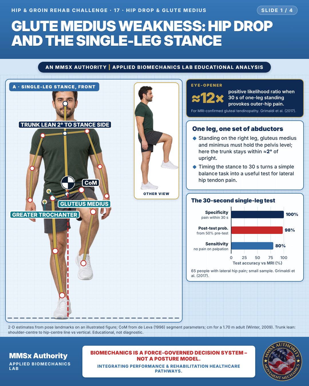
One leg, one set of abductors
Standing on the right leg, gluteus medius and minimus must hold the pelvis level; here the trunk stays within ≈2° of upright.
Timing the stance to 30 s turns a simple balance task into a useful test for lateral hip tendon pain.
The 30-second single-leg test
Movement B: Pelvis dropping (front view)
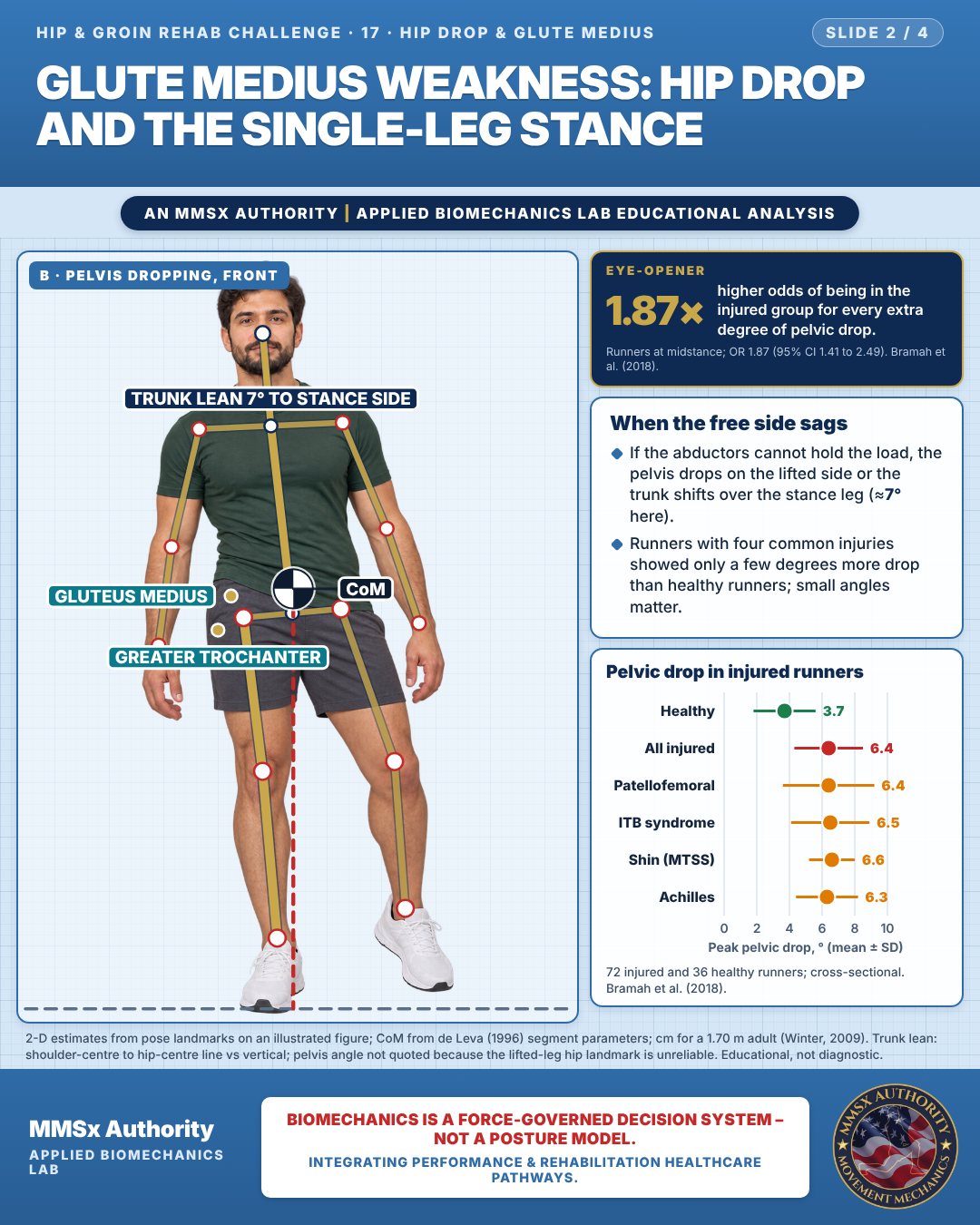
When the free side sags
If the abductors cannot hold the load, the pelvis drops on the lifted side or the trunk shifts over the stance leg (≈7° here).
Runners with four common injuries showed only a few degrees more drop than healthy runners; small angles matter.
Pelvic drop in injured runners
Movement C: Side-lying abduction
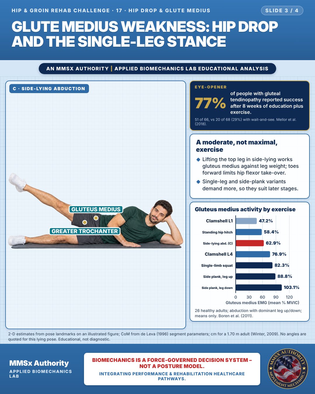
A moderate, not maximal, exercise
Lifting the top leg in side-lying works gluteus medius against leg weight; toes forward limits hip flexor take-over.
Single-leg and side-plank variants demand more, so they suit later stages.
Gluteus medius activity by exercise
Decision pathway
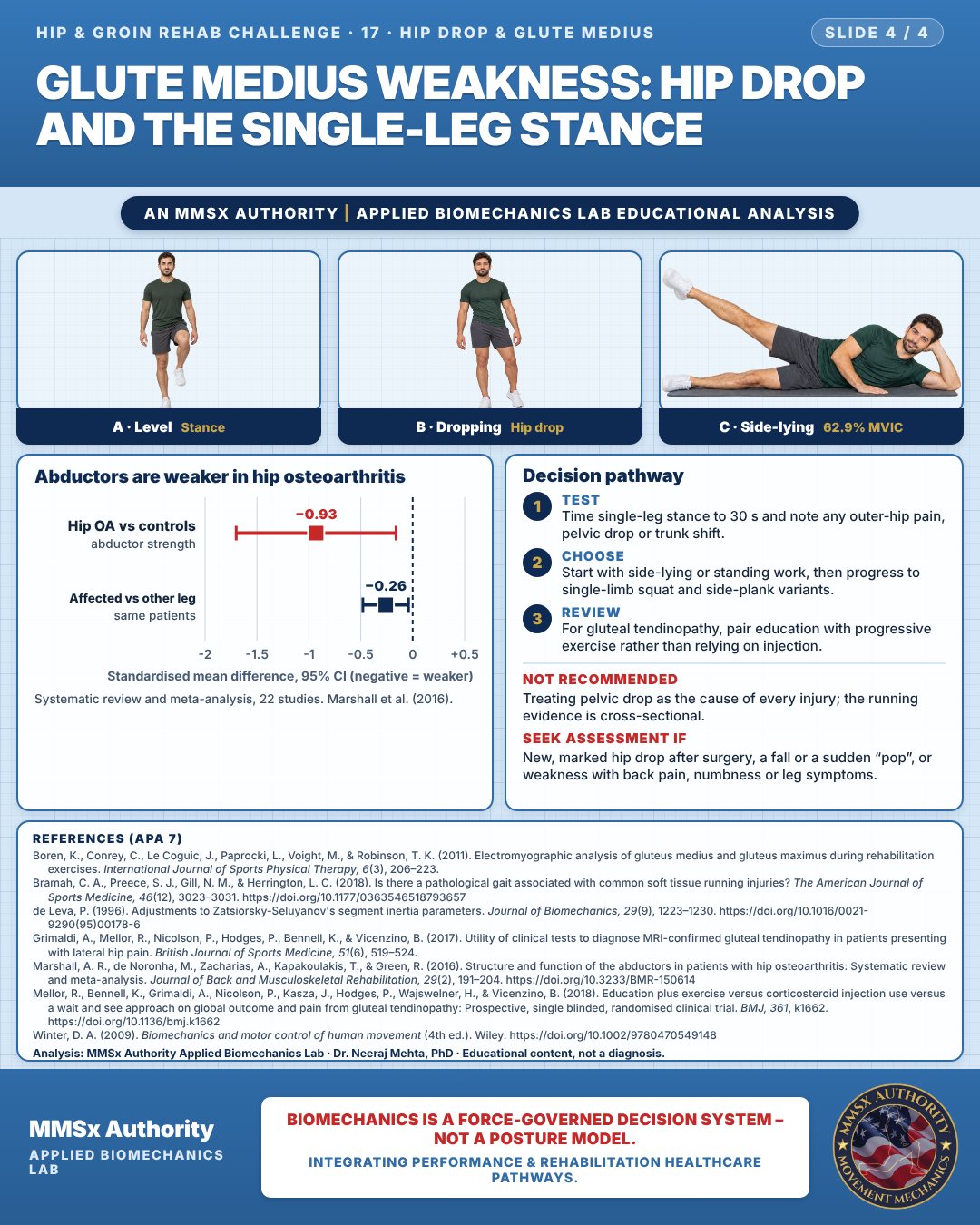
Abductors are weaker in hip osteoarthritis
Test
Time single-leg stance to 30 s and note any outer-hip pain, pelvic drop or trunk shift.
Choose
Start with side-lying or standing work, then progress to single-limb squat and side-plank variants.
Review
For gluteal tendinopathy, pair education with progressive exercise rather than relying on injection.
What the evidence supports
- Choose exercises by load: weight-bearing and side-plank variants activate gluteus medius more than side-lying abduction (Boren et al., 2011).
- Not recommended: treating pelvic drop as the cause of every injury; the evidence is cross-sectional (Bramah et al., 2018).
Myth and evidence
Myth“Side-lying leg raises are the best glute medius exercise.”
EvidenceIn the same participants, side-lying abduction reached 62.9% MVIC, while the single-limb squat reached 82.3% and side-plank variants 88.8–103.1% (Boren et al., 2011).
Cautions
Not recommended
Treating pelvic drop as the cause of every injury; the running evidence is cross-sectional.
Seek assessment if
New, marked hip drop after surgery, a fall or a sudden “pop”, or weakness with back pain, numbness or leg symptoms.
Methods note
2-D estimates from pose landmarks on an illustrated figure; CoM from de Leva (1996) segment parameters; cm for a 1.70 m adult (Winter, 2009). Trunk lean: shoulder-centre to hip-centre line vs vertical. Trunk lean: shoulder-centre to hip-centre line vs vertical; pelvis angle not quoted because the lifted-leg hip landmark is unreliable. No angles are quoted for this lying pose. These are educational estimates, not a diagnosis.
References
- Boren, K., Conrey, C., Le Coguic, J., Paprocki, L., Voight, M., & Robinson, T. K. (2011). Electromyographic analysis of gluteus medius and gluteus maximus during rehabilitation exercises. International Journal of Sports Physical Therapy, 6(3), 206–223.
- Bramah, C. A., Preece, S. J., Gill, N. M., & Herrington, L. C. (2018). Is there a pathological gait associated with common soft tissue running injuries? The American Journal of Sports Medicine, 46(12), 3023–3031. https://doi.org/10.1177/0363546518793657
- de Leva, P. (1996). Adjustments to Zatsiorsky-Seluyanov's segment inertia parameters. Journal of Biomechanics, 29(9), 1223–1230. https://doi.org/10.1016/0021-9290(95)00178-6
- Grimaldi, A., Mellor, R., Nicolson, P., Hodges, P., Bennell, K., & Vicenzino, B. (2017). Utility of clinical tests to diagnose MRI-confirmed gluteal tendinopathy in patients presenting with lateral hip pain. British Journal of Sports Medicine, 51(6), 519–524.
- Marshall, A. R., de Noronha, M., Zacharias, A., Kapakoulakis, T., & Green, R. (2016). Structure and function of the abductors in patients with hip osteoarthritis: Systematic review and meta-analysis. Journal of Back and Musculoskeletal Rehabilitation, 29(2), 191–204. https://doi.org/10.3233/BMR-150614
- Mellor, R., Bennell, K., Grimaldi, A., Nicolson, P., Kasza, J., Hodges, P., Wajswelner, H., & Vicenzino, B. (2018). Education plus exercise versus corticosteroid injection use versus a wait and see approach on global outcome and pain from gluteal tendinopathy: Prospective, single blinded, randomised clinical trial. BMJ, 361, k1662. https://doi.org/10.1136/bmj.k1662
- Winter, D. A. (2009). Biomechanics and motor control of human movement (4th ed.). Wiley. https://doi.org/10.1002/9780470549148
Cite this article
Mehta, N. (2026, October 28). Glute medius weakness: hip drop and the single-leg stance. MMSx Authority Applied Biomechanics Lab. https://mmsxauthority.org/edu/analysis/hip-groin/17-glute-medius-weakness-hip-drop-and-the-single-leg-stance/
Part of the collection
Mehta, N. (2026). Hip and Groin Rehabilitation: 30 Evidence-Based Biomechanics Analyses [Collection]. MMSx Authority Applied Biomechanics Lab. Zenodo. https://doi.org/10.5281/zenodo.23735806
All Hip & Groin Rehab Challenge articles · Rehabilitation Library